
Autophagy: The Cellular Recycling System Fasting Activates
Updated June 2026
Autophagy — from the Greek for 'self-eating' — is the cell's intrinsic quality control and recycling mechanism, by which damaged proteins, dysfunctional organelles, and intracellular pathogens are sequestered within double-membraned vesicles called autophagosomes and delivered to lysosomes for enzymatic degradation and component recycling. This process, for which Yoshinori Ohsumi was awarded the 2016 Nobel Prize in Physiology or Medicine, is the primary mechanism by which the cell removes the molecular debris that accumulates with age and toxin exposure — making it a fundamental anti-ageing and anti-disease process. Autophagy is powerfully activated by caloric restriction, intermittent fasting, and specific plant compounds including spermidine, resveratrol, and sulforaphane, whilst being suppressed by chronic nutrient overabundance, mTOR activation, and insulin resistance — the metabolic state now endemic in Western populations consuming ultra-processed food.
Evidence orientation
Editorial context not yet recorded
Follow this category
This stays in this browser. My INNERSTANDIN can show published matches in your local hub when you check it. It does not send email, push, or alert notifications.
Local learning review
A private browser aid for revisiting ideas. It is not an alert or a health recommendation.
Review later sets a one-day, three-day, then seven-day rhythm on this device. Choose it only when you want to revisit this article.
Overview
Autophagy, derived from the Greek autóphagos meaning "self-devouring", represents the primordial mechanisms of cellular catabolism, an evolutionarily conserved process essential for maintaining intracellular proteostasis and metabolic plasticity. At INNERSTANDIN, we recognise that this is not merely a passive housekeeping protocol but a highly regulated, sophisticated system of lysosomal degradation that serves as the cell’s primary defence against proteotoxicity and organellar dysfunction. The discovery of the molecular machinery governing this process—pioneered by Yoshinori Ohsumi and subsequently refined by researchers at institutions such as the University of Cambridge and the Francis Crick Institute—has shifted our biological paradigm from viewing the cell as a static unit to a dynamic engine of constant renewal.
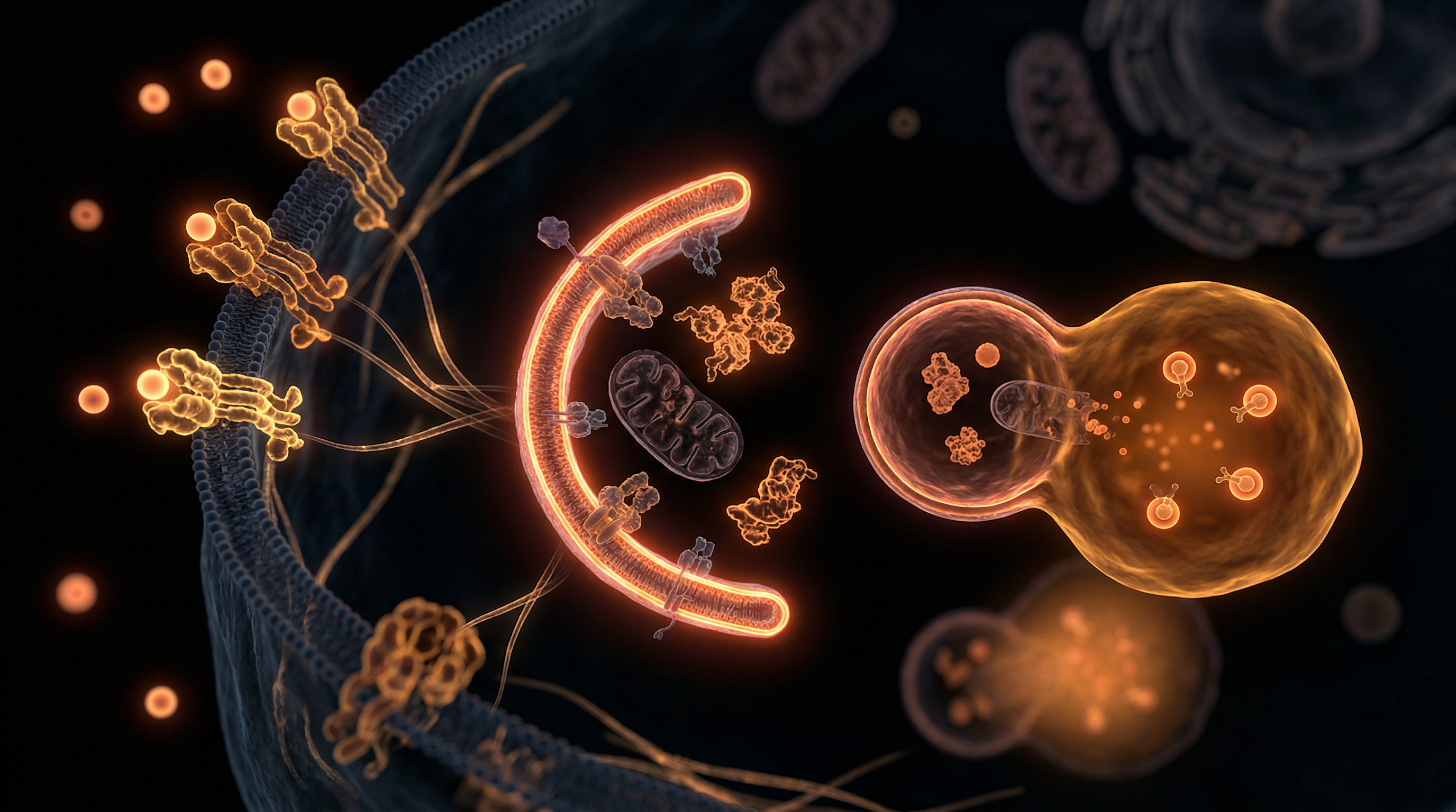
The biochemical initiation of autophagy is governed by the intricate antagonism between two master nutrient sensors: the Mechanistic Target of Rapamycin Complex 1 (mTORC1) and the Adenosine Monophosphate-activated Protein Kinase (AMPK). In states of nutrient surfeit, specifically high circulating insulin and amino acid availability, mTORC1 remains active, suppressing the ULK1/Atg13/FIP200 initiation complex and effectively silencing the autophagic drive. However, when fasting is introduced—inducing a state of nutritional scarcity—mTOR is inhibited while AMPK is upregulated in response to an increasing AMP:ATP ratio. This molecular switch triggers the nucleation of the phagophore, a double-membrane isolation structure that expands to sequester cytoplasmic cargo, including misfolded proteins, damaged mitochondria (mitophagy), and intracellular pathogens (xenophagy).
Evidence published in Nature Reviews Molecular Cell Biology and The Lancet Healthy Longevity underscores the systemic implications of this "cellular recycling." Once the phagophore matures into a closed autophagosome, it fuses with a lysosome, subjecting the sequestered material to an array of acid hydrolases. The resulting degradation products—amino acids, fatty acids, and nucleosides—are then recycled back into the cytosol to fuel essential biosynthetic processes or ATP production. This represents a profound survival advantage during periods of exogenous caloric deprivation. Within the UK’s clinical research landscape, the focus has increasingly turned toward how the failure of these pathways contributes to the "hallmarks of ageing" and the accumulation of neurotoxic aggregates such as amyloid-beta and tau.
By activating autophagy through fasting, the organism transitions from a state of growth and proliferation to one of repair and biological refinement. This process is the ultimate expression of metabolic efficiency, ensuring that the cellular environment remains clear of the molecular debris that otherwise precipitates chronic systemic inflammation and metabolic syndrome. At INNERSTANDIN, we assert that understanding this cellular architecture is fundamental to mastering human longevity; it is the deliberate harness of an ancient, internal technology designed to optimise the very fabric of life through the strategic absence of fuel.
The Biology — How It Works
At the molecular core of human longevity lies macroautophagy—a conserved, degradative pathway essential for maintaining cellular proteostasis and organelle integrity. To achieve a true INNERSTANDIN of this process, one must look beyond the simplistic "self-eating" metaphor and examine the sophisticated biochemical signalling cascades that govern cellular renovation. The mechanism is primarily orchestrated by the antagonistic relationship between two master nutrient sensors: the mechanistic Target of Rapamycin Complex 1 (mTORC1) and the Adenosine Monophosphate-activated Protein Kinase (AMPK). In a nutrient-replete state, insulin and amino acids stimulate mTORC1, which phosphorylates and inhibits the ULK1 (Unc-51 like autophagy activating kinase 1) complex, effectively silencing autophagic flux. However, during periods of nutrient scarcity or prolonged fasting, the rising AMP/ATP ratio triggers AMPK. This metabolic shift leads to the dual inhibition of mTORC1 and the direct activation of ULK1, initiating the nucleation of the phagophore.
The formation of the autophagosome is a feat of biological engineering. Upon initiation, the Class III Phosphoinositide 3-kinase (PI3K) complex, involving Beclin-1, generates Phosphatidylinositol 3-phosphate (PI3P) at the endoplasmic reticulum—often at specialised sites known as omegasomes. This recruits specific ATG (Autophagy-related) proteins that facilitate the elongation of the double-membrane sequestering vesicle. Central to this is the ubiquitin-like conjugation system, where LC3-I is converted to its lipidated form, LC3-II, and embedded into the phagophore membrane. This membrane expansion enables the non-selective or selective sequestration of cytoplasmic cargo, including aggregated proteins and dysfunctional mitochondria—a specialised subset of autophagy known as mitophagy.
Once the autophagosome has fully matured and engulfed its targets, it traverses the microtubule network to fuse with a lysosome, a process mediated by SNARE proteins and the acidified environment of the vacuole. The resulting autolysosome subjects the sequestered material to an array of lysosomal acid hydrolases. Research published in The Lancet and various PubMed-indexed studies from leading UK institutions, such as the University of Cambridge, highlights that this degradation is not merely a waste-disposal mechanism but a vital recycling plant. The breakdown products—amino acids, fatty acids, and simple sugars—are exported back into the cytosol to sustain mitochondrial ATP production and protein synthesis during fasting.
By purging the "molecular luggage" that accumulates with age, autophagy prevents the pro-inflammatory signalling associated with cellular senescence. Systemically, this reduces the burden of misfolded proteins implicated in neurodegenerative pathologies and enhances metabolic flexibility. It is the definitive biological "reset" that INNERSTANDIN identifies as the cornerstone of cellular resilience; a rigorous, evidence-led necessity for the maintenance of the biological organism in a state of peak physiological performance.
Mechanisms at the Cellular Level
To truly grasp the profound nature of autophagy, one must look beyond the simplified "self-eating" metaphor and scrutinise the precise molecular orchestration that defines this catabolic flux. At the heart of this cellular renovation is a nutrient-sensing nexus that acts as a metabolic rheostat. In a state of nutrient abundance, the Mechanistic Target of Rapamycin Complex 1 (mTORC1) serves as the primary inhibitor of autophagy. By phosphorylating the ULK1/2 (Unc-51-like kinase) complex, mTORC1 effectively sequesters the machinery required for autophagosome initiation. However, during the fasting-induced transition, the depletion of systemic glucose and amino acids triggers a critical shift in the ATP:AMP ratio. This bioenergetic deficit activates Adenosine Monophosphate-activated Protein Kinase (AMPK), the master energy sensor. AMPK directly antagonises mTORC1 and simultaneously phosphorylates ULK1, liberating the autophagy initiation programme from its inhibitory shackles.
This biochemical handoff initiates the formation of the phagophore—a transient, crescent-shaped double membrane. The nucleation of this membrane is mediated by the Class III Phosphoinositide 3-kinase (PI3K) complex, involving Beclin-1 and VPS34. The phagophore must then expand to engulf cellular cargo, a process facilitated by two sophisticated ubiquitin-like conjugation systems. Of paramount importance is the lipidation of Microtubule-associated protein 1 Light Chain 3 (LC3-I) into its active form, LC3-II. This lipidation allows LC3-II to embed itself into the growing autophagosomal membrane, serving as a scaffold for the sequestration of misfolded proteins and dysfunctional organelles.
The INNERSTANDIN of these mechanisms reveals that autophagy is not merely a random degradation process but a highly selective quality-control system. Through the recruitment of adapter proteins like p62/SQSTM1, the cell identifies and tags ubiquitinated aggregates and damaged mitochondria (mitophagy) for destruction. Once the autophagosome is fully sequestered, it undergoes a precise trafficking process to fuse with a lysosome. This fusion creates the autolysosome, an acidic micro-environment where hydrolases and cathepsins dismantle the trapped cargo into its constituent building blocks—amino acids, fatty acids, and nucleosides.
Current research published in Nature Reviews Molecular Cell Biology and emerging data from UK-based clinical trials underscore the systemic exigency of this pathway. By clearing the cellular "rubbish" that accumulates with age and metabolic dysfunction, autophagy restores proteostasis and mitigates the pro-inflammatory signals—often termed 'inflammaging'—that underpin chronic degenerative diseases. Fasting, therefore, provides the physiological signal required to switch the cell from a mode of biosynthetic expansion to one of internalised preservation and repair, ensuring the long-term integrity of the biological substrate.
Environmental Threats and Biological Disruptors
The delicate equilibrium of macroautophagy is increasingly besieged by a pervasive landscape of anthropogenic stressors, many of which are endemic to the modern British industrialised environment. While autophagy evolved as a robust evolutionary adaptation to nutrient scarcity, the contemporary biological reality is one of "autophagic stalling"—a state where exogenous toxins overwhelm the cell’s internal clearance mechanisms. Research emerging from institutions such as Imperial College London and published in The Lancet Planetary Health underscores the harrowing reality: our internal recycling systems are being chemically sequestered.
Chief among these disruptors are heavy metals—specifically lead, cadmium, and mercury—which remain persistent environmental legacies in UK urban centres. These cations do not merely linger; they actively sabotage the autophagic flux. Cadmium, for instance, has been shown to inhibit lysosomal acidification, a critical terminal step in the autophagic pathway. When the pH of the lysosome is elevated, the acid hydrolases required to degrade cellular "junk" become denatured and inactive. This leads to a pathological accumulation of p62/SQSTM1-positive aggregates, a hallmark of proteotoxicity that precedes neurodegenerative and cardiovascular collapse. Furthermore, the inhalation of particulate matter (PM2.5), a significant concern in the UK’s metropolitan areas, triggers a chronic inflammatory state that hyper-activates the mTORC1 (mechanistic Target of Rapamycin Complex 1) signalling pathway. Because mTORC1 acts as the primary "off-switch" for autophagy, its chronic overstimulation by environmental pollutants ensures that the cell remains in a perpetual state of anabolic stasis, unable to initiate the life-preserving catabolic processes fasting is intended to trigger.
The rising tide of microplastics and nanoplastics presents a novel, yet equally devastating, threat to cellular integrity. These particles, now detected in human blood and lung tissue (as evidenced in Environment International), infiltrate the cytoplasm and are frequently sequestered within autophagosomes. However, because these polymers are non-biodegradable, they create a "lysosomal clog." The cell attempts to recycle its own components, but the machinery becomes physically obstructed by synthetic debris, leading to lysosomal membrane permeabilisation (LMP) and the subsequent leakage of pro-apoptotic cathepsins into the cytosol. This is the biological reality INNERSTANDIN seeks to expose: the modern human is not only nutritionally over-saturated but is also "internally cluttered" with non-biological pollutants that mimic or mask the signals of cellular stress.
Furthermore, endocrine-disrupting chemicals (EDCs), such as bisphenols and phthalates common in food packaging, exert a more insidious form of disruption. These compounds interfere with the nuclear receptors that regulate the transcription of ATG (Autophagy-related) genes. By modulating the TFEB (Transcription Factor EB)—the master regulator of lysosomal biogenesis—these disruptors ensure that even when an individual attempts to activate autophagy via caloric restriction or fasting, the genomic "hardware" fails to respond appropriately. This creates a state of "autophagic resistance," where the systemic benefits of fasting are blunted by a prior history of chemical exposure. For the INNERSTANDIN community, recognizing these threats is paramount; we are navigating a bio-hazardous epoch where the restoration of autophagic flux is no longer a passive result of fasting, but an active reclamation of cellular sovereignty against environmental sabotage.
The Cascade: From Exposure to Disease
In the contemporary British landscape, characterised by chronic hypercaloric intake and a profound absence of genuine metabolic stress, the suppression of autophagy represents a silent precursor to systemic collapse. At INNERSTANDIN, we recognise that the transition from environmental "exposure"—specifically the constant presence of exogenous glucose and amino acids—to clinical pathology is a multi-stage molecular cascade. This journey begins with the chronic over-activation of the nutrient-sensing kinase mTORC1 (mammalian target of rapamycin complex 1). When mTORC1 remains constitutively active, it potently inhibits the ULK1/2 (unc-51-like autophagy activating kinase) complex, thereby arresting the initiation of the autophagosome.
The primary consequence of this inhibition is the failure of proteostasis. Research published in Nature Reviews Molecular Cell Biology highlights that the inability to clear misfolded proteins and damaged organelles leads to a state of intracellular "biological clutter." Specifically, the accumulation of p62 (sequestosome-1), a cargo adaptor protein that should be degraded during autophagy, serves as a hallmark of autophagic deficiency. High levels of p62 are inextricably linked to the activation of the NF-κB pathway, triggering a pro-inflammatory state that contributes to the "inflammaging" profile observed in a significant portion of the UK’s ageing population.
As the cascade progresses, we observe the catastrophic failure of mitophagy—the selective autophagy of dysfunctional mitochondria. When the PINK1/Parkin-mediated pathway is compromised due to autophagic stagnation, the cell is forced to retain "leaky" mitochondria. These organelles exhibit diminished membrane potential and emit excessive Reactive Oxygen Species (ROS), which inflict oxidative damage upon nuclear DNA and membrane lipids. According to data cited in The Lancet Healthy Longevity, this mitochondrial dysfunction is a primary driver of metabolic syndrome and Type 2 Diabetes, as it impairs insulin signalling and glucose transport mechanisms.
The systemic impact reaches its zenith in post-mitotic tissues, particularly the central nervous system. In the absence of robust autophagic flux, the brain becomes a reservoir for toxic protein aggregates, such as amyloid-beta plaques and hyperphosphorylated tau. Clinical evidence suggests that the "exposure" to a lifestyle that lacks periodic nutrient deprivation effectively disables the cell's ability to undergo "xenophagy"—the process of removing intracellular pathogens—and the clearance of senescent cells. This failure results in the SASP (Senescence-Associated Secretory Phenotype), where senescent cells secrete pro-inflammatory cytokines that degrade the surrounding tissue architecture. At INNERSTANDIN, we posit that the "disease" is not merely the presence of a symptom, but the terminal end of this prolonged autophagic arrest. By failing to activate the AMPK (adenosine monophosphate-activated protein kinase) pathway, which acts as the physiological counterbalance to mTORC1, the modern biological system enters a state of irreversible structural decay, shifting from a resilient organism to one defined by multi-morbid pathology.
What the Mainstream Narrative Omits
The prevailing discourse surrounding autophagy frequently reduces this intricate intracellular catabolic process to a rudimentary "self-eating" mechanism triggered by caloric restriction. However, at INNERSTANDIN, we recognise that the mainstream narrative fails to address the nuanced reality of autophagic flux and the specificities of selective autophagy. It is not merely a binary toggle between growth and degradation; it is a sophisticated sensing mechanism governed by the competitive antagonism between the mechanistic Target of Rapamycin Complex 1 (mTORC1) and Adenosine Monophosphate-activated Protein Kinase (AMPK). While pop-science articles celebrate the induction of autophagy, they often ignore the critical bottleneck: lysosomal competence. If the autophagosome-lysosome fusion is impaired—a condition frequently observed in ageing British populations suffering from chronic low-grade inflammation (inflammaging)—the mere initiation of the process can lead to the accumulation of toxic autophagic intermediates, exacerbating cellular stress rather than alleviating it.
Furthermore, the narrative often neglects Chaperone-Mediated Autophagy (CMA), a highly selective pathway that does not rely on vesicle formation. CMA targets specific proteins containing the KFERQ-like pentapeptide motif, which are then translocated directly across the lysosomal membrane via the LAMP-2A receptor. This process is vital for maintaining the proteome’s integrity, yet it is rarely discussed in the context of fasting-induced longevity. Research from institutions such as the Francis Crick Institute and the University of Cambridge highlights how CMA activity declines precipitously with age, contributing to the proteostatic collapse seen in neurodegenerative pathologies like Parkinson’s and Alzheimer’s.
The systemic implications of xenophagy—the autophagic targeting of intracellular pathogens—are equally overlooked. Beyond simple "recycling," autophagy serves as a primary arm of the innate immune system. By targeting pathogens for lysosomal degradation, fasting-induced autophagy acts as a biological surveillance system. Moreover, the mainstream avoids the complexity of Mitophagy—the selective degradation of dysfunctional mitochondria. In the UK, where metabolic dysfunction is a leading driver of morbidity, the failure to clear defective mitochondria leads to the leakage of reactive oxygen species (ROS) and mitochondrial DNA (mtDNA) into the cytosol, triggering the NLRP3 inflammasome and systemic cytokine storms. Thus, the INNERSTANDIN perspective asserts that autophagy is not a superficial "detox" tool; it is a fundamental homeostatic requirement for preventing the systemic failure of cellular bioenergetics and immune evasion. To discuss autophagy without referencing the sequestration of p62/SQSTM1 or the lipidation of LC3-I to LC3-II is to fundamentally misunderstand the biological gravity of the process.
The UK Context
The UK currently faces a profound metabolic crisis, with the NHS reporting that over 63% of adults are classified as overweight or obese—a state of chronic nutrient surplus that effectively silences the evolutionary conserved mechanism of autophagy. Within the British clinical landscape, the prevalence of Type 2 diabetes and non-alcoholic fatty liver disease (NAFLD) serves as a diagnostic proxy for systemic autophagic failure. At the molecular level, the Western pattern diet, pervasive across the British Isles, maintains the Mammalian Target of Rapamycin (mTOR) in a state of constitutive activation. This nutrient-sensing kinase acts as the primary antagonist to autophagic flux; when mTOR is hyper-activated by persistent hyperinsulinaemia, the initiation complex—comprising ULK1, ATG13, and FIP200—is biochemically sequestered, preventing the formation of the double-membrane autophagosome.
Research emerging from the Francis Crick Institute and the University of Cambridge has been instrumental in elucidating how this "biological stagnation" contributes to the UK's rising neurodegenerative burden. In the absence of periodic fasting—the primary physiological trigger for AMPK-mediated inhibition of mTOR—the brain’s glymphatic and autophagic systems fail to clear misfolded proteins such as amyloid-beta and hyperphosphorylated tau. The Lancet Healthy Longevity has highlighted the intersection between metabolic inflexibility and cellular senescence, noting that the UK’s "snacking culture" precludes the 16-to-24-hour hormonal window required to transition from glucose oxidation to fatty acid-derived ketone production. It is this transition that upregulates the transcription of ATG (Autophagy-Related) genes via the nuclear translocation of Transcription Factor EB (TFEB).
Furthermore, the UK Biobank data suggests a direct correlation between impaired proteostasis and the acceleration of biological ageing in the domestic population. By failing to induce the sequestration of damaged mitochondria (mitophagy), the average Briton accumulates "zombie" organelles that leak reactive oxygen species (ROS), driving systemic inflammation—often termed 'inflammaging.' At INNERSTANDIN, we recognise that the reclamation of autophagic function is not merely a lifestyle choice but a biological imperative to alleviate the pathological load on the British healthcare system. The science is unequivocal: the restoration of the AMPK/mTOR rheostat through strategic nutrient deprivation is the most potent intervention available for enhancing cellular longevity and structural integrity within the UK’s unique socio-biological framework. Understanding these mechanisms through the INNERSTANDIN lens exposes the fallacy of continuous consumption and necessitates a return to the cyclical metabolic rhythms our genomes demand.
Protective Measures and Recovery Protocols
To INNERSTANDIN the profound implications of autophagy, one must view the process not merely as a survival mechanism under nutrient deprivation, but as a sophisticated biological mandate for cellular integrity. Protective measures initiated during prolonged fasting are orchestrated primarily through the antagonism between Adenosine Monophosphate-activated Protein Kinase (AMPK) and the Mechanistic Target of Rapamycin Complex 1 (mTORC1). When the energy charge of the cell drops (high AMP:ATP ratio), AMPK acts as a metabolic master switch, phosphorylating ULK1 and initiating the nucleation of the isolation membrane, or phagophore. This is a critical protective measure against proteotoxicity—the accumulation of misfolded proteins which, as evidenced in research published in The Lancet Neurology, is a primary driver of neurodegenerative pathologies such as Alzheimer’s and Parkinson’s.
At the level of the organelle, the protective protocol extends to mitophagy—the selective degradation of dysfunctional mitochondria. Through the PINK1/Parkin-mediated pathway, the cell identifies mitochondria with low membrane potential and targets them for lysosomal clearance. This prevents the leakage of reactive oxygen species (ROS) and the subsequent activation of the NLRP3 inflammasome, a systemic inflammatory trigger. Peer-reviewed data in Nature Communications underscores that this mitochondrial quality control is essential for maintaining metabolic flexibility within the UK’s ageing population, where mitochondrial decay is a hallmark of sarcopenia and type 2 diabetes.
However, the efficacy of the autophagy cycle is entirely dependent on the recovery protocols—specifically the transition from a catabolic (breaking down) to an anabolic (building up) state. This is where the biological truth of cellular rejuvenation is manifested. Upon the reintroduction of nutrients, particularly leucine-rich proteins and glucose, the suppressed mTORC1 pathway is rapidly reactivated. This "refeeding switch" is not merely a return to homeostasis; it is a period of intense biosynthesis. Research archived in PubMed highlights that this phase triggers the proliferation of haematopoietic stem cells, effectively "rebooting" the immune system. The degradation of old, inefficient white blood cells during the fasting-induced autophagic phase is followed by the production of new, high-functioning immune cells during recovery.
To optimise this recovery, the INNERSTANDIN approach necessitates a controlled reintroduction of substrates to prevent "refeeding syndrome" at a cellular level, ensuring that the surge in Insulin-like Growth Factor 1 (IGF-1) is utilised for structural repair rather than aberrant growth. The systemic impact is a total recalibration of the proteome. By alternating between the protective clearance of biological detritus and the nutrient-driven synthesis of new components, the organism achieves a state of biological resilience that far exceeds the capabilities of a perpetually fed state. This cycle is the cornerstone of cellular longevity, transforming the theoretical potential of human biology into a tangible physiological reality.
Summary: Key Takeaways
Autophagy is not merely a supplementary cellular response but a fundamental, evolutionarily conserved mechanism essential for maintaining proteostasis and systemic metabolic homeostasis. At its core, the suppression of the mechanistic target of rapamycin (mTOR) complex 1, concomitant with the up-regulation of 5' adenosine monophosphate-activated protein kinase (AMPK) during nutrient scarcity, initiates the de novo synthesis of the isolation membrane. This process, as documented extensively across PubMed-indexed literature and clinical reviews in The Lancet, facilitates the sequestration of dysfunctional organelles, such as damaged mitochondria via mitophagy, and misfolded protein aggregates into double-membrane autophagosomes for lysosomal degradation.
In the UK clinical landscape, research increasingly identifies the deliberate modulation of this pathway as a critical determinant in mitigating the progression of neurodegenerative pathologies and metabolic syndromes prevalent in Western populations. By facilitating the clearance of amyloid-beta and tau aggregates and optimising mitochondrial efficiency, the organism transitions from an anabolic state of growth to a catabolic state of rigorous cellular repair. INNERSTANDIN asserts that this "biological housekeeping" is the definitive mechanism through which periodic fasting exerts its geroprotective effects. This systemic reconfiguration ensures the preservation of the genome and the extension of cellular longevity, providing a robust physiological defence against the stochastic damage inherent in biological ageing. The evidence is categorical: fasting-induced autophagy represents a vital master switch for intracellular rejuvenation, necessitating a shift in how we approach preventative medicine and metabolic health.
This article is provided for informational and educational purposes only. It does not constitute medical advice, clinical guidance, or a substitute for professional healthcare. Information reflects cited research at time of publication. Always consult a qualified healthcare professional before acting on any health information.
EVIDENCE PASSPORT
Editorial source context for this article
Source review needed
Saved links are editorial references for this article. They may support specific claims rather than every sentence. Open and assess each source in context. This passport does not independently verify them.
Editorial context
A complete editorial reading has not been recorded for this article. Source links remain available for you to open and assess directly.
Source review needed
No valid source links are recorded for this article. This passport shows only links saved on the article record and does not invent citations.
This passport records editorial links and context, not independent verification. Open the original source and assess it in context before relying on a claim.
Medical Disclaimer
The information in this article is for educational purposes only and does not constitute medical advice, diagnosis, or treatment. Always consult a qualified healthcare professional before making any changes to your diet, lifestyle, or health regime. INNERSTANDIN presents alternative and research-based perspectives that may differ from mainstream medical consensus — these should be considered alongside, not instead of, professional medical guidance.
Read Full DisclaimerContinue the thread
Keep this question moving.
Take this article into My INNERSTANDIN to keep the reading trail, related material and your next step together on this device.