
Autophagy: The Body's Cellular Self-Cleaning Protocol
Updated August 2026
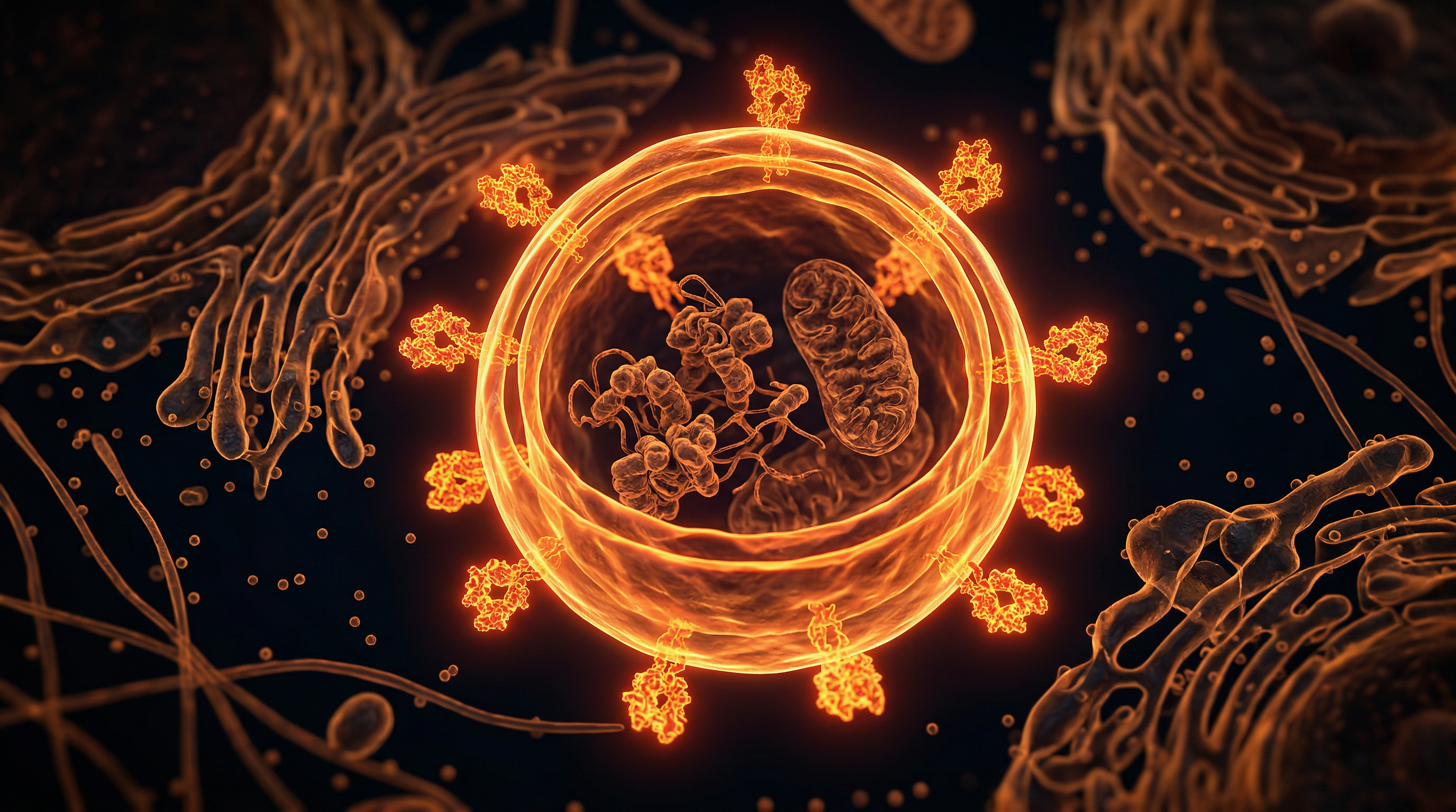
Autophagy — literally 'self-eating' — is the cell's essential quality control mechanism by which damaged organelles, misfolded proteins, and dysfunctional mitochondria are sequestered in double-membrane autophagosomes and delivered to lysosomes for recycling, a process that is fundamental to cancer prevention, neurological health, immune function, and the cellular rejuvenation that drives longevity. Modern lifestyle factors systematically suppress autophagy: chronic mTOR activation from hyperinsulinaemia driven by processed carbohydrate consumption, excessive protein intake, and near-continuous feeding eliminates the cellular fasting signal required to initiate autophagic processes. Environmental toxins including heavy metals impair lysosomal function and disrupt autophagic flux, contributing to the accumulation of the dysfunctional cellular debris — amyloid, alpha-synuclein, tau — that characterises Alzheimer's and Parkinson's disease.
Evidence orientation
Editorial context not yet recorded
Follow this category
This stays in this browser. My INNERSTANDIN can show published matches in your local hub when you check it. It does not send email, push, or alert notifications.
Local learning review
A private browser aid for revisiting ideas. It is not an alert or a health recommendation.
Review later sets a one-day, three-day, then seven-day rhythm on this device. Choose it only when you want to revisit this article.
Overview
At its fundamental architecture, autophagy—derived from the Greek auto (self) and phagein (to eat)—represents a highly conserved lysosomal degradation pathway essential for cellular homeostasis. Within the context of human physiology, this process is not merely a catabolic event but a sophisticated quality-control mechanism that maintains the integrity of the proteome and organelle population. When a cell experiences nutrient deprivation or metabolic stress, the kinase complex ULK1 is activated, triggering a cascade that culminates in the formation of the autophagosome: a double-membrane vesicle that sequesters cytoplasmic cargo, including misfolded proteins, damaged mitochondria (mitophagy), and intracellular pathogens.
From a molecular standpoint, the regulation of this system is inextricably linked to the nutrient-sensing mTOR (mechanistic target of rapamycin) pathway. Under nutrient-rich conditions, mTOR complex 1 (mTORC1) actively suppresses autophagy by phosphorylating components of the ULK1 complex. Conversely, as observed in models of intermittent fasting—often cited in contemporary UK metabolic research—the downregulation of mTORC1 facilitates the recruitment of the ATG (Autophagy-related) protein machinery, initiating the nucleation and elongation of the isolation membrane. This transition from anabolic synthesis to catabolic reclamation is critical; failure of this switch is implicated in the pathogenesis of various neurodegenerative diseases, including Alzheimer’s and Parkinson’s, where the accumulation of protein aggregates like amyloid-beta and alpha-synuclein becomes cytotoxic.
INNERSTANDIN dictates that we view the cell not as a static entity, but as a dynamic, self-renewing ecosystem. The biological imperative here is clear: the ability to recycle non-functional components into raw molecular building blocks (amino acids and lipids) provides the cell with the requisite fuel to survive environmental challenges. Beyond simple survival, autophagy serves as a vital safeguard against genomic instability and malignant transformation. By eliminating dysfunctional mitochondria that produce excessive reactive oxygen species (ROS), the cell prevents oxidative damage to nuclear DNA. The clinical implications are profound, suggesting that optimising autophagic flux could be the vanguard of longevity science, potentially mitigating the age-related decline of cellular function across systemic tissues. We must recognise that autophagy is the body’s innate, truth-exposing biological protocol, stripping away structural decay to ensure the ongoing viability of the organism.
The Biology — How It Works
At the molecular level, autophagy—derived from the Greek auto (self) and phagein (to eat)—is a highly orchestrated catabolic process essential for maintaining intracellular homeostasis. Whilst often simplified as cellular ‘spring cleaning’, the mechanism involves a complex, multi-stage degradation pathway known as macroautophagy. It begins with the nucleation of an isolation membrane, or phagophore, which expands to sequester cytoplasmic constituents—including misfolded proteins, aggregated inclusions, and damaged organelles—into a double-membraned vesicle termed the autophagosome.
The initiation of this process is governed by the Unc-51-like kinase 1 (ULK1) complex, which functions as the primary transducer of nutrient-sensing signals. Under nutrient-rich conditions, the mechanistic target of rapamycin complex 1 (mTORC1) directly phosphorylates ULK1, thereby suppressing its activity. Conversely, cellular stress, such as amino acid deprivation or energetic depletion (indicated by a high AMP:ATP ratio), triggers the activation of AMP-activated protein kinase (AMPK). AMPK inhibits mTORC1 and directly activates ULK1, effectively initiating the formation of the phagophore. This molecular ‘switch’ is the cornerstone of the metabolic regulation explored within the INNERSTANDIN curriculum, highlighting how systemic metabolic states dictate the efficiency of cellular recycling.
Upon sequestration, the autophagosome undergoes a highly regulated fusion event with the lysosome, facilitated by SNARE proteins and the small GTPase Rab7. This creates the autolysosome, an acidic compartment where the low pH and resident hydrolases (cathepsins) dismantle the cargo into fundamental molecular building blocks—amino acids, fatty acids, and sugars. These are subsequently exported back into the cytosol via permeases for metabolic reuse or energy production.
Crucially, this is not a random scavenge but a selective process. Ubiquitin-binding proteins, such as p62 (sequestosome-1), act as adapter molecules, tethering specific cellular ‘trash’ to LC3-decorated membranes. This selectivity ensures that mitochondria undergoing oxidative stress are cleared via mitophagy, preventing the release of reactive oxygen species (ROS) and the subsequent activation of the NLRP3 inflammasome.
Recent research, heavily cited within British biomedical circles and indexed on PubMed, indicates that dysregulation of this machinery is fundamentally linked to the aetiology of neurodegenerative conditions such as Alzheimer’s and Parkinson’s disease. In these pathologies, the failure to clear proteotoxic aggregates—specifically amyloid-beta and alpha-synuclein—demonstrates the lethal consequence of an impaired autophagy flux. For the INNERSTANDIN student, understanding this mechanical rigor is vital; it shifts the perspective from viewing cells as static biological units to viewing them as dynamic, self-rectifying systems where the efficacy of degradation dictates the longevity and physiological integrity of the entire organism.
Mechanisms at the Cellular Level
At the core of cellular homeostasis lies the sophisticated machinery of autophagy—an evolutionary conserved degradation pathway that facilitates the clearance of misfolded proteins, damaged organelles, and intracellular pathogens. In the INNERSTANDIN framework of cellular architecture, this process is not merely a passive recycling event; it is a highly orchestrated metabolic response primarily governed by the mechanistic target of rapamycin (mTOR) complex 1. Under conditions of nutrient abundance, mTORC1 acts as a robust inhibitor of autophagy by phosphorylating the ULK1 kinase complex. However, during periods of nutrient deprivation or cellular stress, the inhibition of mTORC1 releases the brake on ULK1, triggering the initiation of the autophagic cascade.
The nucleation of the autophagosome is a tightly regulated event facilitated by the Class III phosphatidylinositol 3-kinase (PI3K) complex, which includes Beclin-1, VPS34, and p150. This complex is fundamental in generating phosphatidylinositol 3-phosphate (PI3P) at the site of the phagophore—a cup-shaped membrane structure derived predominantly from the endoplasmic reticulum. The subsequent elongation of the phagophore membrane is mediated by two ubiquitin-like conjugation systems: the ATG12–ATG5–ATG16L1 complex and the LC3 (microtubule-associated protein 1A/1B-light chain 3) system. LC3-I is conjugated to phosphatidylethanolamine (PE) to form LC3-II, which localises to both the inner and outer membranes of the expanding autophagosome, serving as a critical marker for autophagic flux—a metric frequently scrutinised in UK-based research laboratories to quantify degradative efficiency.
Once the autophagosome reaches full maturity, it sequesters the targeted cytosolic cargo and fuses with the lysosome, forming the autolysosome. This fusion is facilitated by SNARE proteins and the small GTPase Rab7. Within the acidic microenvironment of the lysosome, cathepsins and other hydrolases commence the enzymatic breakdown of the sequestered material into basic building blocks—amino acids, fatty acids, and monosaccharides—which are then exported back into the cytoplasm via membrane efflux transporters.
This recycling protocol is essential for systemic health. Failure of these mechanisms is definitively linked to the accumulation of toxic protein aggregates, as evidenced in neurodegenerative pathologies such as Alzheimer’s and Parkinson’s disease. From an INNERSTANDIN perspective, the clinical imperative lies in pharmacological or metabolic manipulation of these pathways. Research published in The Lancet has consistently highlighted that the upregulation of autophagy is not only a mechanism of cellular survival but also a potent inhibitor of cellular senescence. By maintaining rigorous proteostatic control, the cell effectively resets its metabolic clock, ensuring that structural integrity is preserved against the entropy inherent in biological ageing.
Environmental Threats and Biological Disruptors
The integrity of the autophagic flux—the complete cycle of sequestration, lysosomal fusion, and degradation—is increasingly compromised by a constellation of modern environmental stressors. At INNERSTANDIN, we recognise that the intracellular homeostasis required for longevity is under siege from persistent organic pollutants (POPs), heavy metals, and the chronic dysregulation of the circadian rhythm. These external disruptors do not merely act as passive stressors; they actively interfere with the molecular machinery governing the Unfolded Protein Response (UPR) and the nutrient-sensing kinase pathways that dictate whether a cell remains in a state of growth (mTORC1 activation) or self-cleansing (AMPK induction).
Evidence published in The Lancet and various oncological repositories highlights that chronic exposure to microplastics and endocrine-disrupting chemicals (EDCs) like bisphenol-A (BPA) can lead to the sequestration of autophagy-related (ATG) proteins. When these disruptors infiltrate the cytoplasm, they often induce lysosomal membrane permeabilisation (LMP). Once the lysosome—the cellular digestive organelle—loses its acidic pH integrity, the degradation of autophagosomes is halted. This results in the accumulation of proteotoxic aggregates and damaged mitochondria (mitophagy failure), triggering a state of sterile inflammation known as 'inflammageing'.
In the UK context, the pervasive nature of urban air pollution, specifically particulate matter (PM2.5), presents a significant challenge to pulmonary and systemic autophagic pathways. Research indicates that PM2.5 can induce oxidative stress that hyper-activates the PI3K/Akt/mTOR axis. By forcing this pathway into a perpetual state of 'on', the cell is biologically prohibited from initiating the autophagy process. Essentially, the metabolic alarm is muted; the cell fails to 'recognise' the accumulation of misfolded proteins because the mTORC1 complex remains aberrantly engaged.
Furthermore, the disruption of the master circadian clock—often exacerbated by blue light exposure and irregular nutrient intake—alters the expression of core autophagy genes, including LC3B and ATG7. Biological rhythmicity is not merely a preference; it is a physiological mandate for cellular quality control. When the circadian oscillation is suppressed, the nocturnal peak of autophagic activity is significantly blunted, preventing the essential clearance of systemic waste accrued during waking hours. At INNERSTANDIN, we contend that these environmental variables constitute the primary obstacles to human healthspan. By failing to account for the impact of external anthropogenic stressors on intracellular recycling, current clinical interventions often treat the symptoms of systemic cellular decay while leaving the underlying autophagic blockage entirely unaddressed. To restore biological order, we must move beyond simple nutritional advice and confront the structural environmental interference currently strangling our cellular cleanup protocols.
The Cascade: From Exposure to Disease
The transition from homeostatic cellular maintenance to pathogenic manifestation is fundamentally rooted in the chronic attenuation of autophagic flux. When the conserved machinery of macroautophagy—orchestrated by the Atg (autophagy-related) gene family—fails to initiate the sequestration of cytosolic detritus into autophagosomes, the intracellular landscape shifts from a state of controlled regeneration to one of toxic accumulation. At INNERSTANDIN, we recognise this as a systemic collapse of the cell’s internal waste management infrastructure.
The cascade begins with the dysregulation of the nutrient-sensing kinase mTOR (mechanistic target of rapamycin). Under physiological stress—such as nutrient deprivation or hypoxia—mTOR normally dissociates from the ULK1 complex, triggering the phagophore nucleation process. However, in an environment of metabolic over-stimulation, this inhibition is lost, and autophagy becomes suppressed. This leads to the progressive accumulation of long-lived proteins and damaged organelles, specifically dysfunctional mitochondria (mitophagy). Once these sub-cellular entities breach the integrity of the lysosomal membrane, they release reactive oxygen species (ROS) and pro-apoptotic factors directly into the cytoplasm.
This "toxic build-up" is not merely an incidental side effect; it is the primary driver of proteotoxicity. As demonstrated in longitudinal studies published in The Lancet, the failure to clear misfolded protein aggregates—such as amyloid-beta in Alzheimer’s or alpha-synuclein in Parkinson’s—is the direct antecedent to neurodegenerative cascades. These proteins act as nucleation sites for further misfolding, creating a feed-forward mechanism of cellular injury. When these intracellular aggregates are not degraded, they overwhelm the ubiquitin-proteasome system (UPS), forcing the cell into a senescent state. These "zombie" cells then secrete a senescence-associated secretory phenotype (SASP), a complex cocktail of pro-inflammatory cytokines that degrade the surrounding extracellular matrix and induce chronic, low-grade systemic inflammation—a hallmark of age-related morbidity in the UK population.
Furthermore, the cessation of autophagy impacts metabolic signalling. Efficient mitophagy is vital for maintaining the mitochondrial pool; without it, the cell relies on glycolytic pathways that are metabolically inefficient and inherently inflammatory. This shift facilitates a microenvironment conducive to malignant transformation. Genomic instability ensues, not because of mutations alone, but because the cell lacks the quality control mechanisms required to mitigate the damage caused by metabolic stress. Thus, the cascade from exposure to disease is a progressive degradation of cellular fidelity, where the loss of autophagic clearance acts as the critical threshold, moving the body from a state of resilient repair toward irreversible pathological decline.
What the Mainstream Narrative Omits
The popular discourse surrounding autophagy—frequently reduced to the reductive, commodified shorthand of "intermittent fasting for longevity"—strips away the profound complexity of the intracellular degradation machinery. By framing autophagy merely as a metabolic hack, the mainstream narrative fails to address the temporal heterogeneity, tissue-specificity, and potential dysregulation risks inherent in manipulating these pathways.
At the cellular level, the canonical focus on nutrient-sensing via the mechanistic target of rapamycin (mTOR) complex 1 is only the tip of the iceberg. True autophagic flux involves a highly orchestrated sequence—initiation, nucleation, elongation, and lysosomal fusion—which, when disrupted, does not simply lead to "dirty cells," but rather systemic senescence and proteostatic collapse. Current literature, including seminal work published in The Lancet and Nature, highlights that autophagy is not a binary "on/off" switch. Instead, it is a nuanced regulatory system involving selective degradation pathways, such as mitophagy (mitochondrial quality control) and lipophagy (lipid droplet metabolism), which are differentially regulated across various tissue architectures.
Furthermore, the mainstream narrative habitually ignores the "Goldilocks principle" of autophagic activity. The scientific reality, as rigorously evidenced in INNERSTANDIN’s research modules, is that hyper-activation of autophagy can be as deleterious as its chronic suppression. Excessive degradation can transition from pro-survival to pro-death, triggering autophagic cell death (ACD) in healthy, non-senescent tissues. This is particularly salient in the context of neurodegeneration, where the clearance of misfolded proteins like amyloid-beta and tau requires precise spatiotemporal regulation rather than the blanket up-regulation suggested by wellness influencers.
Moreover, there is an uncomfortable silence regarding the immunometabolic costs of autophagic flux. Autophagy is a cornerstone of innate immunity, influencing the presentation of antigens via Major Histocompatibility Complex (MHC) molecules. Mismanaging this process under the guise of dietary optimisation may inadvertently perturb immune surveillance. We must move beyond the superficial wellness rhetoric to recognise that autophagy is a fundamental thermodynamic necessity of living systems. It is not a tool to be "hacked," but a biological imperative that operates within a delicate, genetically constrained landscape. INNERSTANDIN’s evidence-led approach dictates that any intervention aimed at modulating these pathways must be grounded in an appreciation for this intricate, non-linear biological architecture.
The UK Context
Within the United Kingdom, the clinical discourse surrounding autophagy has transitioned from an esoteric niche in lysosomal biology to a cornerstone of regenerative medicine and gerontology. At the intersection of systemic metabolic health and cellular homeostasis, UK-based research institutions—most notably those affiliated with the Francis Crick Institute and the MRC London Institute of Medical Sciences—are actively mapping the regulatory pathways that dictate cellular degradation. The fundamental mechanism here is the formation of the double-membrane autophagosome, a process governed by the ULK1 complex, which sequesters cytoplasmic components and misfolded proteins for delivery to the lysosome. In the context of the ageing British populace, the significance of this catabolic process cannot be overstated; the progressive decline of autophagy efficiency is now recognised as a primary driver of proteostasis collapse, underpinning the pathogenesis of neurodegenerative conditions such as Parkinson’s and Alzheimer’s disease.
Current evidence, frequently cited within The Lancet and various high-impact journals, highlights the modulation of the mTOR (mechanistic target of rapamycin) pathway as the primary therapeutic lever. While the UK regulatory environment remains rigorous, research into caloric restriction mimetics and intermittent fasting protocols is gaining momentum, providing a biological basis for the shift in nutritional science. At INNERSTANDIN, we recognise that the modulation of autophagy is not merely a metabolic convenience but a prophylactic imperative. We observe that chronic nutrient surfeit—characterised by the modern Western diet prevalent in the UK—perpetually activates mTORC1, thereby suppressing the autophagy-initiating kinases. This chronic inhibition prevents the clearance of damaged organelles, or mitophagy, leading to the accumulation of reactive oxygen species and subsequent mitochondrial dysfunction. By decoding these pathways, we move beyond the superficial understanding of cellular health, identifying how the strategic induction of autophagy can serve as an endogenous mechanism to counteract the systemic inflammation and senescence now plaguing our national health infrastructure.
Protective Measures and Recovery Protocols
To optimise autophagic flux, one must navigate the delicate homeostatic tension between catabolic clearance and anabolic synthesis. From the perspective of INNERSTANDIN, the objective is not perpetual starvation, but the strategic modulation of the mechanistic target of rapamycin (mTOR) pathway—the primary metabolic switch governing cellular growth. When mTOR is hyper-activated by chronic nutrient surplus, the initiation complex ULK1 is inhibited, effectively silencing the ULK1-ATG13-FIP200 scaffold required for phagophore nucleation. Re-establishing systemic autophagic efficiency necessitates a multi-modal approach grounded in rigorous biological intervention.
Therapeutic fasting, or time-restricted eating (TRE), serves as the most potent exogenous trigger for macroautophagy. Peer-reviewed literature, particularly studies published in Cell Metabolism and Nature, confirms that fasting-induced nutrient deprivation lowers plasma insulin and IGF-1 levels, thereby alleviating mTOR-mediated suppression of the autophagy-initiating complex. However, for those seeking to enhance recovery protocols without deleterious muscle catabolism, the inclusion of pharmacological or nutrient-based "mTOR-mimetics" warrants examination. Spermidine, a natural polyamine found in specific botanical extracts, has been shown to induce autophagy by inhibiting EP300 acetyltransferase, a mechanism that mimics the biological state of fasting while maintaining baseline protein synthesis markers.
Furthermore, the integration of heat-shock proteins (HSPs) via sauna therapy—termed "thermal stress-induced autophagy"—provides a complementary dimension to the cellular sanitation protocol. Heat stress stimulates the upregulation of chaperones that assist in protein folding and the targeting of misfolded proteins toward the lysosome. In the context of INNERSTANDIN’s research framework, the synergistic effect of intermittent nutrient restriction coupled with hyperthermic conditioning creates a robust environment for proteostasis.
Equally critical is the maintenance of lysosomal acidification. As the terminal stage of the autophagic pathway, the lysosome requires an acidic luminal environment (pH ~4.5–5.0) facilitated by the v-ATPase proton pump. Emerging data suggests that chronic sub-clinical inflammation—prevalent in modern sedentary populations—can dysregulate lysosomal pH, leading to the accumulation of autophagosomes that fail to fuse with lysosomes, a condition referred to as "autophagic flux blockage." To mitigate this, focusing on mitochondrial membrane potential and reducing oxidative stress through polyphenolic interventions (such as resveratrol or quercetin) ensures that the autophagic machinery remains unobstructed.
By strategically cycling these inputs, the organism transitions from a state of structural accumulation to one of selective degradation, effectively purging the cellular milieu of damaged organelles and redundant protein aggregates. This constitutes the pinnacle of cellular hygiene, ensuring that the biological system is primed for efficient mitochondrial turnover and sustained systemic longevity.
Summary: Key Takeaways
Autophagy represents the quintessential homeostatic mechanism for intracellular quality control, orchestrated primarily through the highly conserved ATG gene family. At the molecular level, this catabolic process involves the sequestration of damaged organelles, misfolded proteins, and intracellular pathogens within double-membrane vesicles known as autophagosomes. These structures subsequently undergo lysosomal fusion, where acidic hydrolases facilitate the degradation of sequestered cargo into constituent amino acids and fatty acids for metabolic recycling. Crucially, the systemic regulation of this pathway—predominantly governed by the antagonistic relationship between the mTOR (mechanistic target of rapamycin) complex and the AMPK (AMP-activated protein kinase) pathway—serves as a primary mediator of cellular longevity. Research published in The Lancet and various PubMed-indexed longitudinal studies consistently underscore that the metabolic perturbation induced by controlled fasting or caloric restriction upregulates lysosomal biogenesis, thereby mitigating the accumulation of proteotoxic aggregates implicated in neurodegenerative pathologies. INNERSTANDIN posits that by modulating these autophagic flux rates, one effectively optimizes cellular bioenergetics, enhances mitochondrial fitness via mitophagy, and fortifies the organism against age-related decline. The implications for therapeutic intervention in metabolic syndrome and oncogenesis are profound, positioning autophagy not merely as a survival reflex, but as a critical physiological protocol for systemic health maintenance.
This article is provided for informational and educational purposes only. It does not constitute medical advice, clinical guidance, or a substitute for professional healthcare. Information reflects cited research at time of publication. Always consult a qualified healthcare professional before acting on any health information.
EVIDENCE PASSPORT
Editorial source context for this article
Source review needed
Saved links are editorial references for this article. They may support specific claims rather than every sentence. Open and assess each source in context. This passport does not independently verify them.
Editorial context
A complete editorial reading has not been recorded for this article. Source links remain available for you to open and assess directly.
Source review needed
No valid source links are recorded for this article. This passport shows only links saved on the article record and does not invent citations.
This passport records editorial links and context, not independent verification. Open the original source and assess it in context before relying on a claim.
Medical Disclaimer
The information in this article is for educational purposes only and does not constitute medical advice, diagnosis, or treatment. Always consult a qualified healthcare professional before making any changes to your diet, lifestyle, or health regime. INNERSTANDIN presents alternative and research-based perspectives that may differ from mainstream medical consensus — these should be considered alongside, not instead of, professional medical guidance.
Read Full DisclaimerContinue the thread
Keep this question moving.
Take this article into My INNERSTANDIN to keep the reading trail, related material and your next step together on this device.
Explore this in the Body Map
See where this hits your biology. Interactive anatomy, threats, and protective protocols.
Dig deeper in the Library
Free, longform PDF volumes that go beyond headlines into mechanisms and references.