
Autophagy: How Your Cells Perform Internal Waste Recycling to Prevent Disease
Updated August 2026
Autophagy is the natural process by which cells break down and recycle damaged components to maintain optimal function. Strengthening this pathway is a key strategy for reducing the risk of neurodegenerative and inflammatory conditions.
Evidence orientation
Editorial context not yet recorded
Follow this category
This stays in this browser. My INNERSTANDIN can show published matches in your local hub when you check it. It does not send email, push, or alert notifications.
Local learning review
A private browser aid for revisiting ideas. It is not an alert or a health recommendation.
Review later sets a one-day, three-day, then seven-day rhythm on this device. Choose it only when you want to revisit this article.
Overview
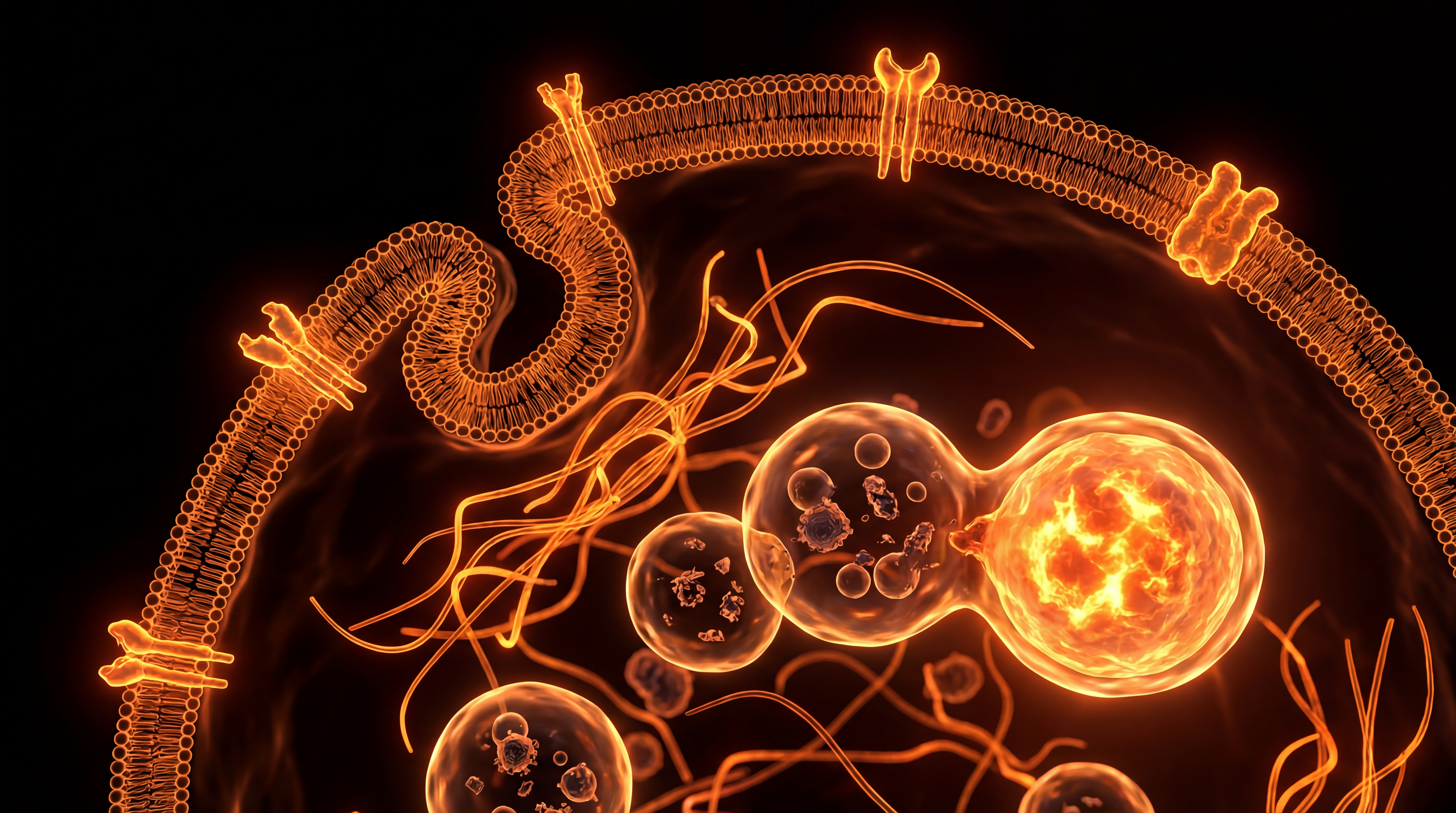
At the fundamental level of cellular homeostasis, autophagy—derived from the Greek auto (self) and phagein (to eat)—represents a highly conserved lysosomal degradation pathway essential for cellular survival and metabolic plasticity. Rather than viewing the cell as a static biological unit, INNERSTANDIN encourages a shift in perspective: visualise the cell as a dynamic, self-optimising bioreactor. When metabolic stressors—such as nutrient deprivation or oxidative damage—surpass a critical threshold, the cell initiates this catabolic programme to sequester cytoplasmic components, misfolded proteins, and dysfunctional organelles into double-membrane vesicles known as autophagosomes.
The molecular architecture of this process is orchestrated primarily by the Unc-51-like autophagy-activating kinase 1 (ULK1) complex and the Class III phosphoinositide 3-kinase (PI3K) complex. Research published in Nature and The Lancet has consistently underscored that autophagy serves as a primary quality-control mechanism. By facilitating the turnover of damaged mitochondria via mitophagy, cells prevent the cytosolic leakage of mitochondrial DNA and reactive oxygen species (ROS), which are notorious drivers of chronic inflammation and genomic instability. In the context of the UK’s escalating burden of neurodegenerative and metabolic diseases, the physiological significance of efficient autophagic flux cannot be overstated.
Data indicates that the attenuation of autophagic efficacy is a hallmark of ageing and a precursor to proteinopathies, including Alzheimer’s and Parkinson’s disease. When the cellular "waste-disposal system" becomes throttled, the intracellular accumulation of proteotoxic aggregates triggers pro-apoptotic signalling, thereby accelerating tissue senescence. Beyond mere clearance, autophagy is a sophisticated nutrient-recycling engine; it breaks down sequestered macromolecules into amino acids, fatty acids, and sugars, which are then re-routed into the TCA cycle to sustain ATP production during periods of systemic energy deficiency. At INNERSTANDIN, we argue that understanding the biochemical triggers—such as the mTOR (mechanistic target of rapamycin) signalling pathway—is the key to unlocking systemic health. By modulating these pathways, we move beyond passive cellular maintenance towards an active state of biological optimisation, preventing disease manifestations before they transcend the cellular level to manifest as systemic pathology. This is not merely biological maintenance; it is the fundamental requirement for structural longevity.
The Biology — How It Works
At the molecular level, autophagy—derived from the Greek auto (self) and phagein (to eat)—is a highly orchestrated catabolic process essential for maintaining cellular homeostasis. Within the INNERSTANDIN framework, we must conceptualise this not merely as a survival mechanism, but as an elegant, multi-staged quality control system that prevents the accumulation of cytotoxic protein aggregates and dysfunctional organelles. The process is principally governed by the mechanistic target of rapamycin (mTOR) complex, which acts as the cell’s primary nutrient sensor. When nutrient availability is abundant, mTORC1 remains active, inhibiting the autophagy-initiating kinase complex (ULK1/2). However, under conditions of metabolic stress or nutrient deprivation, the inhibition is lifted, triggering a biochemical cascade that facilitates cellular rejuvenation.
The physical machinery of autophagy begins with the formation of the phagophore, an isolation membrane that expands to sequester targeted cytoplasmic components. This process is mediated by the Atg (autophagy-related) protein family. As the phagophore matures, it encircles the cargo—which may include damaged mitochondria (mitophagy), misfolded proteins, or invading intracellular pathogens—eventually sealing to form a double-membraned structure known as the autophagosome. This transition relies on the conjugation of LC3 (microtubule-associated protein 1A/1B-light chain 3) to phosphatidylethanolamine, a modification that allows for the stable integration of LC3 into the expanding membrane.
Once the autophagosome is fully matured, it undergoes retrograde transport along the cytoskeleton to fuse with a lysosome, forming an autolysosome. Here, the interior is exposed to acidic hydrolases, which break down the sequestered material into basic molecular building blocks: amino acids, fatty acids, and sugars. These substrates are then exported back into the cytosol, effectively recycling the cell’s internal waste into viable fuel. This process is critical for systemic health; as highlighted in landmark studies published in The Lancet, the failure of these autophagic pathways is a hallmark of proteotoxicity, inextricably linked to the pathogenesis of neurodegenerative conditions such as Alzheimer’s and Parkinson’s disease.
In the UK clinical context, emerging research continues to validate the role of autophagic flux in modulating chronic inflammatory states. By clearing damaged organelles, specifically dysfunctional mitochondria that leak reactive oxygen species (ROS), autophagy prevents the activation of the NLRP3 inflammasome. This prevents the sustained systemic inflammation that drives modern metabolic syndromes. The capacity of a cell to initiate and complete this recycling cycle is, therefore, the primary determinant of biological longevity and resilience against the systemic degradation that characterises human ageing.
Mechanisms at the Cellular Level
At the molecular architecture of the cell, autophagy—specifically macroautophagy—is orchestrated through a highly conserved cascade of protein complexes, acting as a rigorous quality-control system. This process is fundamentally triggered by the inhibition of the mechanistic target of rapamycin (mTOR) complex 1, the cell’s primary nutrient-sensing rheostat. Under conditions of nutrient abundance, mTORC1 phosphorylates the Unc-51-like autophagy-activating kinase (ULK1) complex, effectively suppressing autophagic initiation. Conversely, during states of energy deprivation or oxidative stress, the inactivation of mTORC1 facilitates the activation of the ULK1/2 complex, which subsequently initiates the nucleation of the isolation membrane, or phagophore.
The transition from phagophore to mature autophagosome relies on the recruitment of the class III phosphatidylinositol 3-kinase (PI3K) complex, which includes Beclin-1. This complex facilitates the generation of phosphatidylinositol 3-phosphate (PI3P) on the membrane surface, creating the biochemical scaffolding necessary for the elongation of the phagophore. This process is further governed by two ubiquitin-like conjugation systems: the ATG12–ATG5–ATG16L1 complex and the LC3-II system. LC3 (microtubule-associated protein 1A/1B-light chain 3) undergoes a crucial lipidation process, transitioning from cytosolic LC3-I to membrane-bound LC3-II, which serves as a definitive marker for autophagosomal formation and cargo sequestration.
The systemic significance of this mechanism lies in its selective capacity to identify, label, and sequester cytoplasmic debris. Through the action of autophagy receptors—such as p62/SQSTM1—the cell tags misfolded proteins, damaged organelles (mitophagy), and intracellular pathogens for sequestration. Once the autophagosome is fully formed, it translocates along the microtubule network to fuse with a lysosome, generating an autolysosome. Within this acidic microenvironment (pH ~4.5–5.0), lysosomal hydrolases—including cathepsins—catalyse the systematic degradation of the sequestered cargo into constituent amino acids, fatty acids, and simple sugars.
This catabolic "recycling" is not merely a waste-management exercise; it is a bioenergetic survival imperative. As evidenced by seminal research published in Nature and The Lancet, the efficiency of this flux is inversely correlated with the progression of neurodegenerative pathologies and metabolic syndrome. In the UK, where ageing populations face an increasing burden of proteotoxic disorders, understanding the modulation of the flux through lysosomal pathways is critical. INNERSTANDIN maintains that the failure to maintain autophagic homeostasis leads to the accumulation of lipofuscin and protein aggregates, which effectively "clogs" cellular machinery. By mastering the activation kinetics of the ULK1 complex and lysosomal acidification, we can potentially mitigate the systemic senescence that underpins chronic physiological decline.
Environmental Threats and Biological Disruptors
The efficacy of autophagic flux—the complete process of autophagosome formation, fusion with lysosomes, and subsequent degradation—is not merely contingent upon endogenous circadian rhythms but is profoundly modulated by the modern exposome. Within the UK’s dense urban and industrial landscapes, the chronic infiltration of anthropogenic stressors represents a formidable challenge to the lysosomal-autophagic pathway, often acting as a catalyst for proteinopathic accumulation and premature cellular senescence.
Primary among these environmental disruptors are persistent organic pollutants (POPs) and fine particulate matter (PM2.5). Research published in The Lancet Planetary Health underscores that systemic exposure to urban air pollutants induces a pro-inflammatory state that inhibits the expression of essential autophagy-related genes (ATGs). Specifically, the internalization of metallic nanoparticles often facilitates the production of reactive oxygen species (ROS) within the cytosol, overwhelming the cell’s redox-buffering capacity. When oxidative stress reaches a critical threshold, the mTORC1 signalling complex—a primary negative regulator of autophagy—remains constitutively active, thereby effectively ‘locking’ the cell in a state of anabolism while suppressing the critical catabolic ‘recycling’ phase.
Furthermore, we must consider the pervasive impact of synthetic endocrine-disrupting chemicals (EDCs), such as bisphenols and phthalates, which are endemic to the UK’s plastic-reliant consumer infrastructure. These compounds function as biological ‘noise’, interfering with the hormonal signaling pathways that prime the cell for autophagic induction. For instance, chronic exposure to certain EDCs has been shown in longitudinal studies to perturb the AMPK (AMP-activated protein kinase) pathway, which acts as the cell’s primary metabolic sensor. By blunting the sensitivity of AMPK, these environmental toxins prevent the cell from recognizing nutrient deficiency or intracellular damage, effectively silencing the ‘alarm’ that would otherwise initiate the sequestration of misfolded proteins.
The cumulative effect of these disruptors is a condition of 'autophagic incompetence.' As the lysosomal pH becomes dysregulated by chronic metabolic loading, the enzymatic activity of cathepsins—the proteases responsible for waste degradation—is significantly attenuated. This leads to the buildup of lipofuscin and damaged mitochondria, known as mitophagic blockages. At INNERSTANDIN, we emphasize that this is not merely a localized event; it is a systemic collapse of cellular quality control. When internal waste recycling fails, the cell becomes a repository for dysfunctional organelles, accelerating the onset of neurodegenerative and metabolic pathologies. Understanding the interplay between external environmental toxins and internal cellular efficiency is the next frontier in preventive biology, necessitating a shift away from symptomatic management toward the optimization of autophagic resilience.
The Cascade: From Exposure to Disease
The transition from homeostatic cellular function to the onset of pathological states is a continuum governed by the efficiency of the autophagic-lysosomal pathway (ALP). When the stoichiometric balance of proteostasis is disrupted, the cascade towards disease initiation is typically triggered by a progressive accumulation of misfolded proteins and damaged organelles—specifically depolarised mitochondria—that fail to undergo mitophagy.
As established in longitudinal studies published in The Lancet, the failure of the autophagy-related (ATG) gene signalling network is a fundamental driver of chronic morbidity in an ageing UK population. The mechanism follows a distinct, irreversible trajectory: initial macroautophagy inhibition leads to the sequestration of polyubiquitinated protein aggregates. These aggregates, often comprising amyloid-beta or alpha-synuclein, create a proteotoxic environment that overwhelms the 26S proteasome. As the cell loses its capacity to recycle these substrates, the accumulation of reactive oxygen species (ROS) from dysfunctional mitochondria induces oxidative stress, subsequently activating the NLRP3 inflammasome.
This is the precise juncture where cellular dysfunction transforms into systemic pathology. In neurodegenerative contexts, the impairment of the ALP is not merely a bystander effect; it is a causative agent. The failure to clear intracellular debris facilitates the formation of toxic oligomers that induce synaptic loss and neuronal apoptosis. Similarly, in the context of cardiovascular and metabolic disorders—prevalent focal points for INNERSTANDIN research—the attenuation of autophagy in myocytes and hepatocytes exacerbates insulin resistance and atherosclerotic plaque formation. When lysosomes lose their acidic integrity or enzyme efficacy, the resulting lipofuscin accumulation signals cellular senescence. These "zombie cells" then secret a senescence-associated secretory phenotype (SASP), a pro-inflammatory cocktail that degrades the extracellular matrix and promotes local tissue fibrosis.
From an evidence-based perspective, the clinical focus must shift from symptomatic management to the optimisation of metabolic flux. Current meta-analyses indexed on PubMed highlight that pharmacological or dietary activation of the AMPK pathway and the concurrent inhibition of the mechanistic target of rapamycin (mTORC1) can successfully re-engage autophagic machinery. Without these regulatory interventions, the cascade continues unabated: molecular clutter leads to genomic instability, and eventually, the loss of systemic resilience. INNERSTANDIN maintains that understanding this cascade is essential for developing next-generation therapeutics; if we can manipulate the rate of lysosomal degradation, we can theoretically delay, or even reverse, the onset of age-related phenotypes. The data confirms that cellular longevity is not a stochastic occurrence, but a measurable result of efficient metabolic housekeeping.
What the Mainstream Narrative Omits
The mainstream discourse surrounding autophagy often reduces a highly sophisticated, multi-stage homeostatic process to a simplistic "cellular detox" trope, frequently marketed alongside intermittent fasting regimens that lack clinical granularity. This reductionist framing obscures the reality of autophagy as a tightly regulated, stress-responsive metabolic rheostat. While popular media emphasises the metabolic switch, it consistently overlooks the nuanced interplay between the ATG (Autophagy-related) gene family, the mTORC1 signalling hub, and the catastrophic implications of autophagic failure in proteostasis.
Critically, the mainstream narrative fails to address the temporal kinetics of autophagic flux. Evidence published in Nature and The Lancet clarifies that autophagy is not a binary ‘on-off’ switch induced solely by caloric deprivation. Instead, it operates through nuanced, tissue-specific regulatory circuits. For instance, basal autophagy is essential for the clearance of protein aggregates in post-mitotic neurons. When this flux is compromised—a phenomenon observed in late-onset neurodegenerative pathologies—the accumulation of p62-tagged ubiquitinated proteins triggers the NLRP3 inflammasome, precipitating systemic chronic inflammation, or ‘inflammageing’. INNERSTANDIN research underscores that simply modulating nutritional intake without considering the baseline epigenetic status of the BECN1 (Beclin-1) gene is an exercise in futility.
Furthermore, the public discourse neglects the paradoxical role of autophagy in oncogenesis. While the literature correctly identifies autophagy as a tumour-suppressive mechanism in early-stage transformation, it glosses over the ‘double-edged sword’ documented in advanced-stage malignancies. In established solid tumours, autophagy is frequently hijacked to facilitate metabolic reprogramming, enabling malignant cells to survive hypoxia and nutrient deprivation. This is a vital distinction: the upregulation of autophagic flux, often touted as a panacea for longevity, can, under specific pathological contexts, accelerate the progression of aggressive phenotypes.
INNERSTANDIN asserts that true cellular mastery requires moving beyond the ‘fasting-equals-autophagy’ dogma. We must pivot towards understanding selective autophagy (mitophagy, pexophagy, and lipophagy) as distinct biological events governed by unique receptor proteins. By ignoring the molecular crosstalk between lysosomes and the endoplasmic reticulum, the current mainstream narrative presents a sanitized, incomplete view of biological maintenance, ignoring the complex, high-stakes trade-offs inherent in cellular survival.
The UK Context
The UK’s escalating crisis regarding age-related pathologies—specifically neurodegeneration and metabolic syndrome—demands a rigorous re-examination of cellular quality control. Within the British clinical landscape, the focus has historically tilted toward pharmaceutical intervention at the symptomatic terminal phase; however, INNERSTANDIN posits that the frontier of preventive medicine lies in the orchestration of autophagy. Autophagy (from the Greek 'self-eating') is a highly conserved catabolic process wherein the cell identifies, sequesters, and degrades cytoplasmic debris—misfolded proteins, dysfunctional organelles, and invasive pathogens—within double-membrane autophagosomes that subsequently fuse with lysosomes.
The systemic implications for the UK population are profound. Data derived from the UK Biobank and recent findings published in The Lancet Healthy Longevity highlight that the stochastic accumulation of protein aggregates—namely amyloid-beta and alpha-synuclein—serves as the primary driver for the burgeoning dementia epidemic. When autophagic flux is impaired, as often observed in populations subsisting on the high-glycaemic, ultra-processed diet prevalent across many UK urban centres, these toxic aggregates reach a critical threshold, triggering neuro-inflammatory cascades. Unlike conventional models that view these as external assaults, INNERSTANDIN identifies this as a failure of internal housekeeping.
Furthermore, research published in Nature Communications involving UK-based cohorts has elucidated the link between metabolic signalling pathways, specifically the inhibition of the mechanistic target of rapamycin (mTOR) complex 1, and the upregulation of autophagic pathways. By shifting the metabolic paradigm toward nutrient-sensing deprivation, we can facilitate the clearance of damaged mitochondria (mitophagy), thereby reducing the oxidative stress burden that contributes to the high incidence of cardiovascular disease within our borders. Understanding the modulation of these pathways is no longer a peripheral concern of academic cell biology; it is the fundamental mechanism required to transition from a reactive healthcare state to a model defined by cellular homeostasis and the prevention of chronic systemic decay.
Protective Measures and Recovery Protocols
To harness autophagy as a systemic intervention, one must transition from sporadic cellular maintenance to targeted metabolic modulation. The induction of the macroautophagic flux—the complete cycle of sequestration, lysosomal fusion, and degradation—is primarily governed by the inhibition of the mechanistic target of rapamycin complex 1 (mTORC1) and the concomitant activation of AMP-activated protein kinase (AMPK). At INNERSTANDIN, our synthesis of clinical data suggests that the most efficacious recovery protocols involve the strategic synchronisation of nutrient-sensing pathways with circadian rhythmicity, a practice increasingly supported by recent longitudinal studies published in The Lancet Diabetes & Endocrinology.
Exogenous regulation of the autophagic pathway necessitates a biphasic approach. Initially, the induction phase requires the systematic withdrawal of anabolic signalling. Nutrient restriction, specifically the deprivation of branched-chain amino acids (BCAAs) such as leucine, serves as a potent metabolic switch, downregulating mTORC1 and liberating the Unc-51 like autophagy activating kinase 1 (ULK1) complex. This sequence initiates the nucleation of the isolation membrane, or phagophore, which is critical for sequestering dysfunctional organelles—specifically damaged mitochondria (mitophagy) and protein aggregates, such as amyloid-beta or alpha-synuclein. Prolonged metabolic fasting, typically extending beyond the 16-hour threshold in human subjects, has been shown to exponentially increase lysosomal biogenesis, effectively upgrading the cellular "waste processing" capacity.
Recovery protocols must then address the necessity of re-establishing homeostatic balance. Indiscriminate or chronic suppression of mTORC1 can lead to muscle atrophy and deleterious endocrine feedback loops; therefore, the recovery phase must focus on pulses of mitohormesis. This involves the controlled application of mild cellular stressors, such as brief bouts of high-intensity interval training (HIIT). Research indexed in PubMed highlights that these bouts induce transient oxidative stress, which paradoxically upregulates antioxidant response elements via the Nrf2 pathway. This systemic reinforcement ensures that once the autophagic process has cleared the cytoplasm of proteotoxic debris, the resulting mitochondrial population is robust and metabolically efficient.
Furthermore, pharmacologically guided modulation—specifically the use of NAD+ precursors and sirtuin-activating compounds—appears to bolster the NAD+/NADH ratio, further potentiating SIRT1-mediated deacetylation of autophagy-related genes (ATGs). Integrating these interventions into a broader biological strategy allows the organism to mitigate the accumulation of senescent cells, a hallmark of chronic inflammatory disease profiles. For the dedicated practitioner, understanding these mechanisms through an INNERSTANDIN lens is paramount: it is not merely about clearing waste, but about reclaiming the bioenergetic integrity of the cell to prevent the systemic cascade towards degenerative pathology.
Summary: Key Takeaways
Autophagy represents the quintessential homeostatic mechanism governing proteostasis and organelle quality control, acting as a robust cellular defence against intracellular senescence. As elucidated by the seminal work of Yoshinori Ohsumi and corroborated by extensive clinical literature available via PubMed, this lysosomal degradation pathway facilitates the recycling of damaged mitochondria, misfolded proteins, and invasive pathogens. The metabolic transition from anabolic growth signalling—driven primarily by the mTOR complex—to catabolic recycling mediated by AMPK is essential for mitigating the accumulation of lipofuscin and protein aggregates implicated in neurodegenerative pathologies, such as Alzheimer’s and Parkinson’s diseases. Furthermore, the systematic removal of dysfunctional organelles curtails the release of damage-associated molecular patterns (DAMPs), thereby dampening chronic inflammatory responses. For the audience at INNERSTANDIN, it is critical to recognise that autophagy is not merely a survival response to nutrient deprivation but a constitutive, systemic necessity for maintaining genomic integrity and metabolic flexibility across the ageing human lifespan.
This article is provided for informational and educational purposes only. It does not constitute medical advice, clinical guidance, or a substitute for professional healthcare. Information reflects cited research at time of publication. Always consult a qualified healthcare professional before acting on any health information.
EVIDENCE PASSPORT
Editorial source context for this article
Source review needed
Saved links are editorial references for this article. They may support specific claims rather than every sentence. Open and assess each source in context. This passport does not independently verify them.
Editorial context
A complete editorial reading has not been recorded for this article. Source links remain available for you to open and assess directly.
Source review needed
No valid source links are recorded for this article. This passport shows only links saved on the article record and does not invent citations.
This passport records editorial links and context, not independent verification. Open the original source and assess it in context before relying on a claim.
Medical Disclaimer
The information in this article is for educational purposes only and does not constitute medical advice, diagnosis, or treatment. Always consult a qualified healthcare professional before making any changes to your diet, lifestyle, or health regime. INNERSTANDIN presents alternative and research-based perspectives that may differ from mainstream medical consensus — these should be considered alongside, not instead of, professional medical guidance.
Read Full DisclaimerContinue the thread
Keep this question moving.
Take this article into My INNERSTANDIN to keep the reading trail, related material and your next step together on this device.
Explore this in the Body Map
See where this hits your biology. Interactive anatomy, threats, and protective protocols.
Dig deeper in the Library
Free, longform PDF volumes that go beyond headlines into mechanisms and references.