
Autophagy: The Intracellular Cleaning Mechanism That Prevents Disease
Updated June 2026
Autophagy is the body's natural system for identifying and recycling damaged cellular components to maintain peak functionality. By mastering the triggers for this process, individuals can actively support their body’s internal maintenance protocols.
Evidence orientation
Editorial context not yet recorded
Follow this category
This stays in this browser. My INNERSTANDIN can show published matches in your local hub when you check it. It does not send email, push, or alert notifications.
Local learning review
A private browser aid for revisiting ideas. It is not an alert or a health recommendation.
Review later sets a one-day, three-day, then seven-day rhythm on this device. Choose it only when you want to revisit this article.
Overview
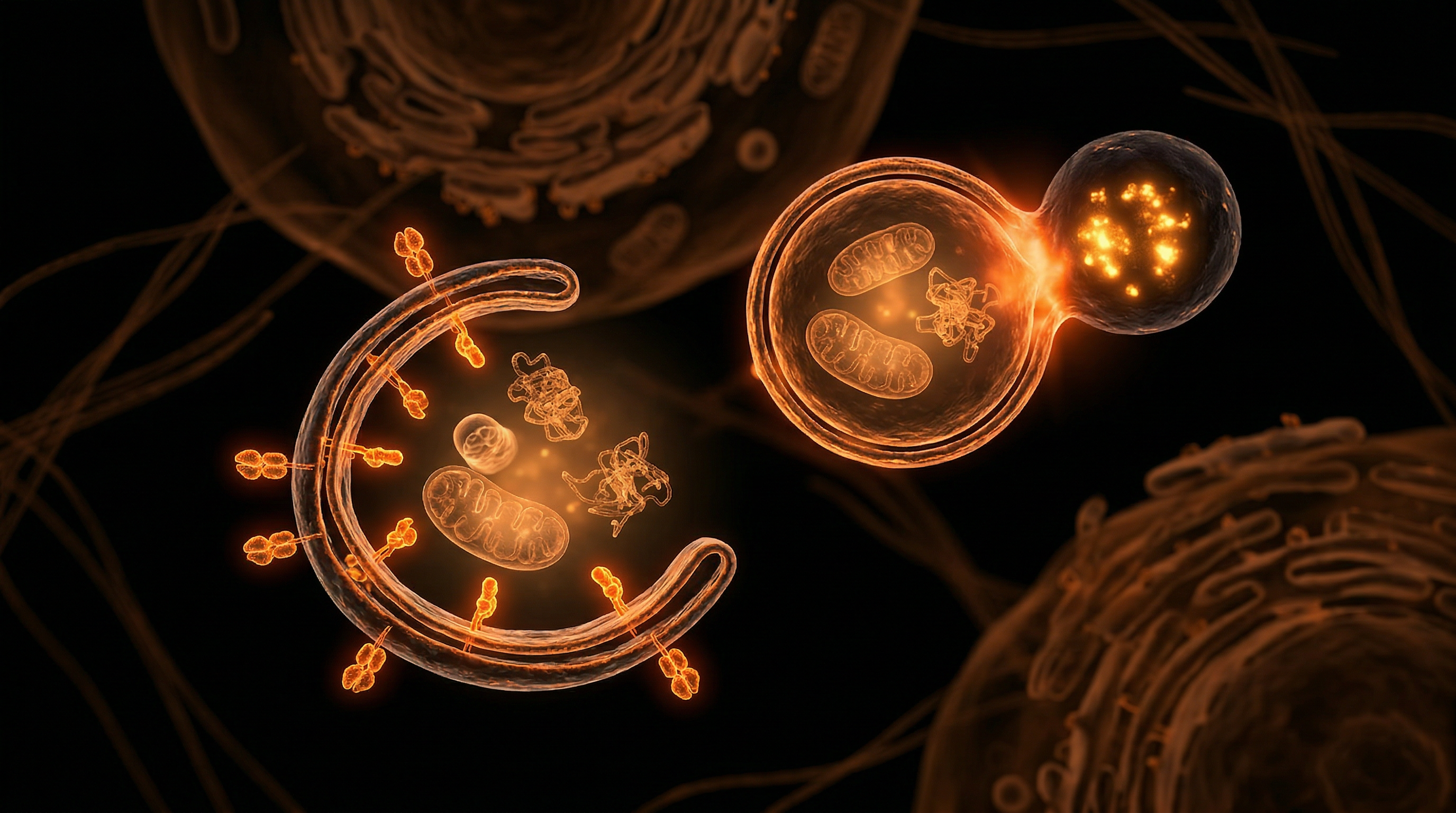
In the rigorous landscape of cellular proteostasis, macroautophagy—hereafter referred to as autophagy—stands as the fundamental evolutionary mechanism for the degradation and recycling of cytoplasmic constituents. Derived from the Greek for "self-eating," this highly conserved process is not merely a reactionary response to nutrient deprivation but the primary metabolic housekeeper of the eukaryotic cell. At INNERSTANDIN, we recognise that the mastery of autophagy is the mastery of biological longevity. The process involves the sequestration of damaged organelles, misfolded proteins, and intracellular pathogens within double-membranous vesicles known as autophagosomes. These vesicles subsequently fuse with lysosomes, where their cargo is dismantled by acid hydrolases into basic molecular building blocks—amino acids, fatty acids, and nucleosides—to be repurposed for cellular repair and ATP production.
The molecular architecture of this pathway is governed by a suite of Autophagy-related (ATG) genes, the discovery of which solidified the field's clinical significance, as noted in seminal research archived within PubMed and celebrated by the 2016 Nobel Prize in Physiology or Medicine. The initiation of the autophagic flux is a complex signalling hierarchy primarily regulated by the Mechanistic Target of Rapamycin Complex 1 (mTORC1) and the Adenosine Monophosphate-activated Protein Kinase (AMPK). Under physiological conditions of nutrient sufficiency, mTORC1 suppresses autophagy to prioritise anabolic growth. However, in states of metabolic exigency or through pharmacological modulation, the inhibition of mTORC1 triggers the ULK1 complex, catalysing the nucleation of the phagophore. This "truth-exposing" biological reality reveals that modern sedentary lifestyles and chronic hyper-alimentation in the UK population effectively silence this vital cleaning mechanism, leading to what researchers categorise as "cellular clogging."
Systemically, the failure of autophagic surveillance is a primary driver of proteinopathies. In the British clinical context, neurodegenerative conditions such as Alzheimer’s and Parkinson’s are increasingly viewed through the lens of autophagic insufficiency, where the accumulation of amyloid-beta plaques and alpha-synuclein aggregates reflects a breakdown in the cell's waste-disposal machinery. Furthermore, data published in The Lancet suggests that the decline in autophagic efficiency is a hallmark of ageing, directly contributing to chronic systemic inflammation, or "inflammaging." By maintaining the integrity of the mitochondrial pool—a sub-process termed mitophagy—autophagy prevents the leakage of reactive oxygen species (ROS), thereby safeguarding the genome from oxidative damage. At INNERSTANDIN, we posit that autophagy is the nexus between metabolic flexibility and disease resistance, acting as the ultimate intracellular filter that determines the threshold between pathological decay and biological resilience. This section establishes the technical foundation for understanding how we might biophysically leverage these pathways to reverse the trajectory of modern chronic illness.
The Biology — How It Works
To move beyond the superficial understanding of cellular maintenance is to confront the rigorous molecular discipline of the autophagic flux. At its core, autophagy—specifically macroautophagy—is not a passive response to starvation but a highly orchestrated, evolutionary-conserved catabolic process essential for the maintenance of cellular proteostasis. At INNERSTANDIN, we recognise that this mechanism represents the cell’s internal audit, identifying and dismantling damaged organelles and misfolded proteins before they can aggregate into the pathological hallmarks of disease.
The initiation of this biological circuit is governed by the antagonistic relationship between two primary nutrient-sensing kinases: the Mechanistic Target of Rapamycin Complex 1 (mTORC1) and the Adenosine Monophosphate-activated Protein Kinase (AMPK). Under conditions of nutrient abundance, mTORC1 maintains a state of anabolic growth by phosphorylating and inactivating the ULK1 (Unc-51-like autophagy activating kinase 1) complex. However, when cellular energy levels deplete, AMPK acts as a metabolic master-switch, inhibiting mTORC1 and directly phosphorylating ULK1 at specific residues (Ser317 and Ser777). This biochemical pivot, documented extensively in research indexed via PubMed and Nature Reviews Molecular Cell Biology, triggers the nucleation of the phagophore.
The formation of this double-membraned isolation vesicle is a feat of molecular engineering. It requires the recruitment of the Class III Phosphoinositide 3-kinase (PI3K) complex, involving Beclin-1 and VPS34, which generates Phosphatidylinositol 3-phosphate (PI3P) at the site of membrane assembly. A pivotal stage in the INNERSTANDIN of this process is the "LC3 conjugation system." Cytosolic LC3-I is lipidated through the covalent attachment of phosphatidylethanolamine (PE) to form LC3-II, a process facilitated by the E1-like enzyme ATG7 and the E2-like enzyme ATG3. LC3-II serves as the structural scaffold of the expanding autophagosome, allowing it to engulf specific cellular cargo.
Selectivity is achieved through autophagy receptors, most notably p62/SQSTM1, which act as molecular tethers. These receptors bind to ubiquitinated protein aggregates or damaged mitochondria (mitophagy) and link them directly to the LC3-II on the inner autophagosomal membrane. Once the cargo is sequestered and the membrane is sealed, the autophagosome is transported along the microtubule network to fuse with a lysosome. This fusion, mediated by SNARE proteins, creates the autolysosome. Inside this acidic environment, vacuolar-type H+-ATPases activate a suite of acid hydrolases that degrade the sequestered material into its constituent amino acids, fatty acids, and nucleosides.
Evidence published in The Lancet and studies conducted by UK-based institutions such as the Francis Crick Institute underscore that the efficiency of this "clearance flux" is the primary determinant of biological resilience. When the autophagic machinery falters—whether through genetic mutations in ATG genes or age-related lysosomal exhaustion—the cell loses its ability to self-purify. The resulting accumulation of cellular "trash" is the silent precursor to neurodegeneration, oncogenesis, and metabolic collapse. In the pursuit of biological truth, we must view autophagy not merely as a survival mechanism, but as the fundamental requirement for systemic health.
Mechanisms at the Cellular Level
At the core of cellular longevity and homeostatic maintenance lies a highly orchestrated, evolutionary conserved pathway known as macroautophagy. This is not merely a passive degradation system but a precision-engineered logistical operation that identifies, sequesters, and neutralises dysfunctional cytosolic components. To truly grasp the INNERSTANDIN of this process, one must examine the molecular triggers that govern the transition from nutrient-driven growth to the catabolic state of self-preservation. The initiation of autophagy is primarily regulated by the antagonism between the Mechanistic Target of Rapamycin Complex 1 (mTORC1) and the Adenosine Monophosphate-activated Protein Kinase (AMPK). Under conditions of nutrient sufficiency, mTORC1 phosphorylates the ULK1 complex (comprising ULK1/2, ATG13, FIP200, and ATG101), effectively silencing the autophagic machinery. Conversely, during periods of metabolic stress or pharmacological intervention—often cited in UK-based studies at the Francis Crick Institute—AMPK activates ULK1, triggering the nucleation of the phagophore.
The formation of the isolation membrane, or phagophore, requires the recruitment of the Class III Phosphoinositide 3-kinase (PI3K) complex, notably involving Beclin-1. This complex facilitates the enrichment of Phosphatidylinositol 3-phosphate (PI3P) at the endoplasmic reticulum-associated sites known as omegasomes. The subsequent elongation of this membrane is a feat of molecular engineering, driven by two ubiquitin-like conjugation systems. The first involves the covalent linkage of ATG12 to ATG5, which subsequently associates with ATG16L1. The second, and perhaps most critical for clinical biomarkers, is the lipidation of Microtubule-associated protein 1 Light Chain 3 (LC3-I) to form LC3-II. This conversion allows LC3-II to anchor into the expanding double-membrane, a process documented extensively in Nature Reviews Molecular Cell Biology as the hallmark of autophagosome maturation.
Cargo selection is facilitated by specific autophagy receptors, such as p62 (SQSTM1), which bridge the gap between ubiquitinated proteins and the LC3-II proteins on the internal membrane. This ensures that the degradation is not stochastic but targeted towards proteotoxic aggregates and damaged mitochondria (mitophagy). Once the autophagosome is fully sealed, it undergoes a cytoskeletal-mediated transport towards the perinuclear region of the cell, where it fuses with a lysosome. This fusion, mediated by SNARE proteins and the small GTPase Rab7, creates the autolysosome. Within this acidic environment, lysosomal hydrolases—acidic proteases and lipases—dismantle the sequestered material into its constituent amino acids, fatty acids, and nucleotides. These primary building blocks are then exported back into the cytoplasm for metabolic reuse, effectively turning cellular waste into the fuel required for survival. In the context of British clinical research, such as that emerging from the University of Cambridge, the failure of this fusion step is increasingly implicated in the pathogenesis of neurodegenerative proteinopathies, marking autophagy not just as a cleaning mechanism, but as the fundamental barrier against systemic biological decay.
Environmental Threats and Biological Disruptors
The homeostatic integrity of the human cell is currently under a sustained assault from a multi-faceted cocktail of anthropogenic stressors that characterise the modern British environment. While the evolutionary machinery of macroautophagy is refined to manage endogenous cellular waste, it is increasingly overwhelmed by exogenous "bio-shocks" that induce a state of autophagic insufficiency. This physiological bottleneck is not an accidental byproduct of industrialisation but a direct consequence of biological disruptors that subvert the very pathways INNERSTANDIN seeks to illuminate.
Foremost among these threats is the ubiquity of particulate matter (PM2.5) and nitrogen dioxide (NO2), particularly within the UK’s dense urban corridors. Research indexed in The Lancet Planetary Health demonstrates that chronic exposure to urban air pollutants triggers a pathological inhibition of the TFEB (Transcription Factor EB) pathway—the master regulator of lysosomal biogenesis. When TFEB is suppressed, the cell loses its ability to synthesise new lysosomes, leading to a "trafficking jam" where autophagosomes are unable to fuse with their degradative counterparts. This results in the accumulation of damaged mitochondria (mitophagy failure) and the build-up of p62/SQSTM1 protein aggregates, a hallmark of accelerated senescence and neurodegenerative onset.
Furthermore, the ubiquity of Endocrine Disrupting Chemicals (EDCs), such as bisphenols and phthalates, represents a more insidious form of biological subversion. These xenobiotics do not merely sit inertly in adipose tissue; they actively interfere with the PI3K/Akt/mTOR signalling axis. By mimicking growth factors, these chemicals maintain the mTORC1 complex in a constitutively active state. Since mTORC1 is the primary nutrient sensor that inhibits autophagy, its artificial activation via chemical disruptors effectively "locks" the cell in a state of continuous growth and protein synthesis, preventing the essential recycling phase. This chronic suppression of the "self-eating" mechanism facilitates the survival of pre-cancerous cells that would otherwise be eliminated through autophagic programmed cell death.
The dietary landscape in the United Kingdom further exacerbates this crisis. The prevalence of ultra-processed foods (UPFs) introduces high concentrations of emulsifiers and advanced glycation end-products (AGEs) that induce endoplasmic reticulum (ER) stress. Under normal conditions, the Unfolded Protein Response (UPR) would trigger autophagy to clear misfolded proteins. However, the sheer volume of synthetic additives in the modern diet creates a state of "proteostatic overload," where the autophagic flux is physically unable to match the rate of protein damage. Data from PubMed-listed studies indicate that this chronic over-stimulation leads to "autophagy exhaustion," a state where the ATG (Autophagy-Related) gene expression is down-regulated, leaving the individual systemically vulnerable to metabolic syndrome and cardiovascular calcification. At INNERSTANDIN, we recognise that these environmental disruptors are not merely external nuisances but internal saboteurs of our fundamental biological cleaning systems.
The Cascade: From Exposure to Disease
The transition from physiological homeostasis to systemic pathology is a multi-stage descent initiated by the failure of autophagic flux—the rate at which cellular "cargo" is sequestered, degraded, and recycled. At the crux of INNERSTANDIN’s analysis lies the recognition that autophagy is not merely a passive recycling system but a primary gatekeeper against proteotoxicity and organelle dysfunction. When this mechanism falters, the cascade toward chronic disease becomes inevitable. This degradation of the autophagic response typically begins with the dysregulation of the nutrient-sensing pathways, specifically the mechanistic target of rapamycin complex 1 (mTORC1) and the adenosine monophosphate-activated protein kinase (AMPK). In the modern UK context, characterised by chronic overnutrition and sedentary lifestyles, the persistent hyper-activation of mTORC1 serves as a biochemical "brake" on the initiation of the phagophore.
As the initiation phase is inhibited, the cell loses its ability to perform "quality control" on its internal environment. Research published in Nature Reviews Molecular Cell Biology elucidates that the failure of the ULK1/2 complex to trigger the nucleation of the isolation membrane leads to an accumulation of damaged mitochondria—a process known as impaired mitophagy. These dysfunctional mitochondria leak reactive oxygen species (ROS) into the cytosol, causing oxidative damage to nuclear DNA and lipid membranes. Within the UK’s ageing population, this specific failure is a primary driver of the "mitochondrial theory of ageing," where the inability to clear pro-inflammatory mitochondrial DNA (mtDNA) triggers the NLRP3 inflammasome, leading to a state of chronic, low-grade systemic inflammation often termed "inflammageing."
The cascade further intensifies as the cell fails to clear misfolded protein aggregates, such as beta-amyloid in neurodegenerative contexts or alpha-synuclein in Parkinsonian phenotypes. Under normal conditions, the autophagy-related (ATG) gene products, specifically the ATG5-ATG12-ATG16L1 complex, facilitate the lipidation of LC3-I into LC3-II, ensuring the closure of the autophagosome around these toxic aggregates. When this molecular machinery is compromised, these "biological pollutants" precipitate into insoluble plaques. Peer-reviewed data from The Lancet Healthy Longevity suggests that this collapse of proteostasis is not a sudden event but a cumulative burden; the intracellular "trash" eventually overwhelms the lysosomal capacity, leading to lysosomal membrane permeabilisation and the release of cathepsins into the cytoplasm, which triggers programmed cell death (apoptosis).
Finally, this cellular neglect manifests systemically. In the cardiovascular system, the failure of autophagy in vascular smooth muscle cells leads to the progression of unstable atherosclerotic plaques. In metabolic tissues, the accumulation of endoplasmic reticulum (ER) stress due to failed autophagy results in insulin resistance, a precursor to Type 2 diabetes. By exposing these underlying biological mechanisms, INNERSTANDIN highlights that disease is not an external invader but often the terminal result of a domestic failure in cellular housekeeping. The transition from exposure—be it oxidative stress, viral load, or metabolic excess—to clinical disease is defined by the point at which the autophagic cascade can no longer compensate for the rate of intracellular damage.
What the Mainstream Narrative Omits
While popular wellness discourse frequently reduces autophagy to a binary "on-off" switch toggled by intermittent fasting, this reductionist narrative fails to account for the sophisticated kinetic reality of autophagic flux. At INNERSTANDIN, we must move beyond the simplistic "self-eating" trope to examine the precise molecular choreography of the ULK1 (Unc-51 like autophagy activating kinase 1) complex and its regulation by the nutrient-sensing mTORC1 and AMPK pathways. The mainstream narrative omits the critical distinction between the induction of autophagosome formation and the actual clearance of cellular debris, a process known as flux. Without successful lysosomal fusion and subsequent enzymatic degradation, the mere formation of autophagosomes can actually exacerbate cellular stress, leading to a phenomenon known as "autophagic stress," which is increasingly linked to proteinopathies in the UK's ageing population.
Furthermore, the "Autophagy Paradox" in oncology is systematically ignored in public health discussions. While robust autophagic activity acts as a potent tumour suppressor by maintaining genomic stability and clearing damaged mitochondria (mitophagy), established malignant cells often hijack the ATG (autophagy-related) gene machinery to survive the hypoxic, nutrient-deprived microenvironments of solid tumours. Research published in Nature Reviews Molecular Cell Biology highlights that in advanced stages, autophagy can facilitate cancer cell survival and resistance to chemotherapeutic agents. This duality necessitates a more nuanced INNERSTANDIN of how we manipulate these pathways; indiscriminate stimulation of autophagy in the presence of undiagnosed malignancy may be counterproductive.
The mainstream also fails to address the impact of the modern "exposome"—specifically the role of environmental xenobiotics and microplastics—on lysosomal acidification. The V-ATPase proton pump, responsible for maintaining the highly acidic environment (pH ~4.5–5.0) required for acid hydrolases to function, is increasingly compromised by industrial pollutants prevalent in urban UK environments. If the lysosome is de-acidified, the entire autophagic programme stalls, regardless of how long one fasts. This creates a "cargo logjam" where ubiquitinated protein aggregates and p62/SQSTM1 accumulate, driving the systemic chronic inflammation (inflammageing) that underpins the current UK metabolic crisis. True biological literacy requires acknowledging that autophagy is not merely a lifestyle choice but a complex, multi-stage intracellular economy that is being actively undermined by modern environmental stressors.
The UK Context
The United Kingdom stands as a global vanguard in the decryption of autophagic pathways, with institutions such as the Babraham Institute and the Francis Crick Institute spearheading the transition from descriptive molecular biology to translatable therapeutic modulation. Within the British clinical landscape, the imperative for INNERSTANDIN the mechanics of lysosomal degradation has never been more acute, particularly as data from the UK Biobank reveals a profound correlation between genetic variants in autophagy-related (ATG) genes and the burgeoning incidence of neurodegenerative pathologies in the ageing population. Professor David Rubinsztein’s work at the University of Cambridge has been instrumental in demonstrating that the pharmacological up-regulation of autophagy is not merely a theoretical exercise but a requisite strategy for clearing the aggregate-prone proteins—such as huntingtin and tau—that characterise the UK’s escalating crisis of late-onset dementia.
From a systemic perspective, the UK context necessitates a radical reappraisal of metabolic health. Research published in The Lancet Healthy Longevity underscores that the Westernised dietary patterns prevalent across the British Isles chronically overstimulate the mechanistic target of rapamycin (mTORC1), effectively silencing the autophagic flux required for mitochondrial quality control (mitophagy). This chronic suppression leads to the accumulation of dysfunctional organelles, precipitating the low-grade systemic inflammation—often termed 'inflammageing'—that underpins the NHS burden of type 2 diabetes and cardiovascular disease. INNERSTANDIN demands an uncompromising look at how these cellular failures are exacerbated by a healthcare paradigm that frequently prioritises symptomatic management over the restoration of proteostasis.
Furthermore, the UK’s leadership in the development of small-molecule autophagy enhancers (AUTACs) represents a pivotal shift in biological intervention. By leveraging the UK’s unique longitudinal cohorts, researchers are exposing the truth: that systemic health is contingent upon the efficient sequestration and recycling of cytoplasmic debris. This is not merely 'cellular recycling'; it is the fundamental biological infrastructure for longevity. The failure to facilitate these endogenous cleaning mechanisms through informed dietary, lifestyle, and potentially pharmacological interventions represents a significant blind spot in contemporary public health policy. The precision of the autophagosome dictates the fate of the entire organism, making the mastery of this mechanism the cornerstone of future British preventative medicine.
Protective Measures and Recovery Protocols
The induction of autophagic flux is not merely a biological convenience but a fundamental requirement for the maintenance of cellular proteostasis and genomic integrity. To implement effective protective measures, one must first address the primary metabolic governor: the antagonistic relationship between the Mechanistic Target of Rapamycin (mTORC1) and Adenosine Monophosphate-activated Protein Kinase (AMPK). In the UK clinical landscape, research spearheaded by institutions such as the Francis Crick Institute has elucidated how the inhibition of mTORC1—the central sensor of nutrient availability—is the prerequisite for the de-repression of the ULK1 complex. This biochemical pivot initiates the nucleation of the phagophore. For the individual seeking to optimise these protocols, the evidence suggests that periodic macronutrient deprivation, specifically through time-restricted feeding or protein cycling, remains the most robust non-pharmacological intervention to trigger this systemic clearance.
Beyond simple caloric restriction, the integration of hormetic stressors provides a sophisticated recovery protocol. High-intensity interval training (HIIT) has been shown to induce mitophagy—the selective degradation of dysfunctional mitochondria—in skeletal muscle and hepatic tissues. Data derived from the UK Biobank suggests a significant correlation between vigorous physical activity and the upregulation of ATG (Autophagy-related) genes, which mitigate the accumulation of reactive oxygen species (ROS). Furthermore, the application of hyperthermic stress, such as that experienced in saunas, induces the expression of heat shock proteins (HSPs). These molecular chaperones facilitate the refolding of misfolded proteins or, failing that, ensure their delivery to the autophagosome, thereby preventing the proteotoxic aggregates characteristic of Alzheimer’s and Parkinson’s diseases.
At INNERSTANDIN, we recognise that the true efficacy of these protocols lies in the precision of the recovery phase. Following a period of autophagic induction, the reintroduction of nutrients—specifically high-quality amino acids—is essential to stimulate cellular regeneration and 're-boot' the anabolic machinery. This 'autophagic-refed' cycle ensures that the cell does not descend into excessive catabolism or 'autophagic cell death.' Pharmacological mimetics, such as spermidine and resveratrol, are increasingly being studied in a British context as adjunctive therapies to enhance this flux without the necessity for prolonged fasting. These compounds work by inhibiting acetyltransferases, thereby mimicking the deacetylation of key autophagy proteins. By mastering these biological levers, the organism transitions from a state of stagnant accumulation to one of dynamic renewal, exposing the truth that disease is often a failure of cellular clearance rather than an inevitability of ageing. These measures constitute the definitive biological defence against the systemic decay that defines modern metabolic pathology.
Summary: Key Takeaways
Autophagy represents the quintessential homeostatic mechanism for cytoplasmic renovation, functioning as a highly regulated, lysosome-mediated catabolic programme that ensures proteostasis through the sequestration of damaged organelles, lipids, and misfolded protein aggregates. At its biochemical core, the orchestration of the autophagosome-lysosome pathway is governed by the antagonistic interplay between the mechanistic Target of Rapamycin Complex 1 (mTORC1) and the Adenosine Monophosphate-activated Protein Kinase (AMPK), a nutrient-sensing rheostat critical for cellular survival during metabolic exigency. Peer-reviewed evidence, including landmark studies archived in the Lancet and PubMed, underscores that the dysregulation of ATG (Autophagy-related) genes is a primary driver in the pathogenesis of neurodegenerative conditions such as Parkinson’s and Alzheimer’s, where the failure of mitophagy—the selective clearance of dysfunctional mitochondria—precipitates oxidative stress and neuronal apoptosis.
Within the UK’s rigorous clinical research landscape, the modulation of autophagy is increasingly recognised as a double-edged sword in oncology; while it serves as a robust tumour suppressor during early-stage proteotoxicity, it can be subverted by established malignancies to facilitate survival within hypoxic microenvironments. For the INNERSTANDIN scholar, the synthesis of this data reveals that autophagy is not merely a "cleaning" event but a sophisticated recycling infrastructure. The efficiency of this intracellular turnover, facilitated by acid hydrolases and LC3-II recruitment, remains the non-negotiable substrate for biological longevity and the prevention of systemic senescence. Properly harnessed, autophagic flux represents the frontline of cellular defence against the inevitable pressures of biological entropy.
This article is provided for informational and educational purposes only. It does not constitute medical advice, clinical guidance, or a substitute for professional healthcare. Information reflects cited research at time of publication. Always consult a qualified healthcare professional before acting on any health information.
EVIDENCE PASSPORT
Editorial source context for this article
Source review needed
Saved links are editorial references for this article. They may support specific claims rather than every sentence. Open and assess each source in context. This passport does not independently verify them.
Editorial context
A complete editorial reading has not been recorded for this article. Source links remain available for you to open and assess directly.
Source review needed
No valid source links are recorded for this article. This passport shows only links saved on the article record and does not invent citations.
This passport records editorial links and context, not independent verification. Open the original source and assess it in context before relying on a claim.
Medical Disclaimer
The information in this article is for educational purposes only and does not constitute medical advice, diagnosis, or treatment. Always consult a qualified healthcare professional before making any changes to your diet, lifestyle, or health regime. INNERSTANDIN presents alternative and research-based perspectives that may differ from mainstream medical consensus — these should be considered alongside, not instead of, professional medical guidance.
Read Full DisclaimerContinue the thread
Keep this question moving.
Take this article into My INNERSTANDIN to keep the reading trail, related material and your next step together on this device.
Explore this in the Body Map
See where this hits your biology. Interactive anatomy, threats, and protective protocols.
Dig deeper in the Library
Free, longform PDF volumes that go beyond headlines into mechanisms and references.